Ferric Nitrilotriacetete Augments 7,12-Dimethylbenz(a) Anthracene-Initiated and Benzoyl Peroxide -Promoted Skin Carcinogenesis
Background: Renal cancer is caused by ferric nitrilotriacetete (Fe-NTA), and little is known about its implications on skin cancer. The effects of Fe-NTA on benzoyl peroxide (BPO) -induced tumor stimulation in 7,12-dimethyl benz(a)anthracene (DMBA)-initiated mouse cutaneous carcinogenesis have been presented in this report. Methods: Fe-NTA was administered topically to Swiss mice, and tumors were induced with DMBA. After that, the animals were given BPO for 40 weeks. The occurrence of tumors was documented. Results: Fe-NTA at a dose of 12 mg iron per mouse induced an increase in tumor occurrence over times as compared to the control (DMBA+ BPO) treated group. Tumors appeared earlier in the Fe-NTA group, with a higher incidence number of tumors. In addition, BPO-mediated lipid peroxide induction and [3H] thymidine uptake were greater in Fe-NTA treated group. Conclusion: We propose that Fe-NTA boosts BPO, tumor-promoting potential, and that Fe-NTA-induced oxidative stress is effective for BPO mediated cutaneous carcinogenesis.
Abbreviations
NTA: Nitrilotriacetic Acid; Fe-NTA: Ferric Nitrilotriacetate; BPO: Benzoyl Peroxide.
Introduction
Nitrilotriacetic acid (NTA) is the aminopolycarboxylic acid. It’s a colorless solid that’s been employed as a chelating agent in hospital and household laundry sterilizers in both developed and developing countries. NTA also generates ferric nitrilotriacetate (Fe-NTA) complex [1, 2] by forming coordination compounds (chelates) with metal ions such as Fe3+. Injection of this iron chelate intraperitoneally (i.p.) to rats and mice promotes lipid peroxidation by producing free radicals [3], eventually leading to renal cell carcinoma, pulmonary metastasis and increased tumor mortality rates [4, 5, 6]. This model also showed increases in malondialdehyde, breakdown of polyunsaturated fatty acid lipid peroxides, α, β-unsaturated hydroxyalkenal modified proteins and 8-oxo-
7,8-dihydro-2’-deoxyguanosine (8-OHdG) [7, 8]. According to our study, also Fe-NTA is involved in liver injury According to our study Fe-NTA which involved in liver injury, being featured [9]. These findings suggest that Fe-NTA exerts its carcinogenesis potential through a variety of mechanisms. According to our investigation, the amount of iron in the body during pregnancy has an important role in the development of skin cancer [10]. In another investigation, we reported that iron overload augments benzoyl peroxide (BPO)-mediated tumor promotion in mouse skin [11]. Thereby, the oxidative capability of Fe-NTA may increase oxidative production and increase skin papilloma. Therefore, the purpose of this study was to investigate the role of Fe-NTA on mice skin papilloma induced by DMBA/ BPO.
Methods
Chemicals
The depilatory cream used in this study was obtained from Tolidaro- Iran, tirichloro acetic acid, perchloric acid and nitric acid were obtained from Merck- Germany. While, Nitrilotriacetic acid, ferric nitrate, BPO, thiobarbituric acid and DMBA were purchased from Sigma-Aldrich Chemical Company. [3H] thymidine was obtained from Amersham (Little Chalfont, UK), and all other chemicals were sourced from various chemical suppliers.
Animals
Female albino Swiss mice, free of pathogens, were obtained from the laboratory animal breeding house at Tabriz University of Medical Sciences. The mice were housed under standard conditions, with access to adequate food and water. For chronic experiments, 20 mice (6 weeks old with body Wt. 22 ±1 g) were used, while for acute experiments, 6 mice were recruited. Two days prior to testing, the dorsal hair of the mice was shaved using a hair removal cream to ensure complete removal. The experiments were conducted following the guidelines for the care and use of laboratory animals (National Institutes of Health Publication No. 85-23, revised 1985) and were approved by the Committee on Animal Research of Tabriz University of Medical Sciences. Every attempt was made to minimize both the number of animals used and the degree of suffering experienced by the animals.
Preparation of Fe-NTA
The Fe-NTA iron solution was prepared following the method of Awai, et al. [12]. In summary, iron nitrate and NTA were dissolved separately in distilled water, and then mixed in a molar ratio of (1:3) Fe-NTA. The resulting solution was adjusted to a pH of 7.4 using sodium bicarbonate. The solution was prepared fresh and used immediately.
Preparation of Tissue
At the end of the designated time period, all mice were euthanized, and the dorsal skin was removed from the body and washed with cold normal saline. The epidermis was then cleaned, and one gram of tissue was homogenized and subjected to subcellular fractionation to obtain post- mitochondrial supernatant for biochemical analysis.
Iron Estimation
In summary, 6 mice were chosen from each group of chronically treated animals and one gram of minced tumor tissue, non-tumor tissue, and skin from the control group were placed in a Chinese dish, and 10 ml of acid digestion mixture (nitric acid) was added. The samples were left overnight and most of the tissue had dissolved by the following day. The mixture was then digested on a hot plate until near dryness. Subsequently, 5 ml of water was added and heated until near boiling. From this solution, 1 ml was diluted with water to make a 10 ml solution and analyzed using an Atomic Absorption Spectrophotometer to determine the concentration of iron. The Fe-NTA group with the highest tumorigenic effects was chosen, and the amount of iron in the tumor and non-tumor parts of the skin was measured. A control group that received only acetone was also used for comparison. The tissue iron concentration was then calculated and expressed as mg/g tissue. To estimate iron levels in skin tumors, complete, uninjured tumors were used.
Test Methods
Four groups of animals, each consisting of six mice, were selected for biochemical investigations. Group I was topically treated with acetone alone and served as the control. Groups III and IV received topical application of Fe- NTA (12 mg iron/mouse) for a period of 15 days. Six hours after Fe-NTA treatment, groups II and IV were treated with a single topical application of BPO (20 mg/200 µl acetone/ mouse). Twelve hours after the treatment with BPO, all animals were euthanized, and their skins were removed for biochemical testing. Four groups of mice, each consisting of 20 animals, were selected for the carcinogenicity tests. Group I served as the control. Groups II, III, and IV received daily topical applications of Fe-NTA at doses of 6, 9, and 12 mg iron per mouse, respectively, for a period of 2 weeks. Nine hours after the last administration of Fe-NTA, all groups of mice, including the control group, received a single topical application of 60 μg of DMBA in 200 μl of acetone. One week after DMBA treatment, the mice received twice-weekly topical applications of 20 mg BPO in 200 µl acetone/mouse for a period of 30 weeks. The mice were then monitored for the occurrence of tumours. To see the effect of any of these chemicals alone, mice groups were individually treated with acetone, DMBA, BPO and Fe-NTA alone. Those groups which alone had no tumours were observed during this period, thus these data are not shown in figures. The basis for the identification of various tumors was done according to an earlier description of O’Connell, et al. [13].
Lipid Peroxidation Test
The lipid peroxidation test was carried out with a slight modification of the method by Wright, et al. [14]. Briefly, 0.5 ml of 10% homogeneous liquid was mixed with 1.5 ml of 0.1 M phosphate buffer and incubated at 37°C with gentle shaking for one hour. Next, 1 ml of 10% trichloroacetic acid solution and 0.67% thiobarbituric acid were added sequentially. The test tubes were heated in hot water for 15 minutes, followed by cooling and centrifugation at 5000 g for 10 minutes. The resulting pink-colored solution was analyzed using a spectrophotometer at a wavelength of 535 nm. The results were expressed as nmol of malondialdehyde formed per hour per mg of tissue at 37°C using a molar extinction coefficient of 1.56 x 10⁵ M-1 cm-1.
[3H] Thymidine Uptake Test
The [3H] thymidine uptake assay in skin DNA was conducted following Smart, et al. [15] method. Firstly, a 10% (w/v) homogenate of epidermis was prepared in cold water containing an equal volume of cold TCA (10%). The precipitate was then washed with 5% cold TCA and subsequently incubated with 10% cold perchloric acid for 8 hours. The mixture was centrifuged, and the washing process was repeated with 5% cold perchloric acid. The precipitate was dissolved in 10% warm perchloric acid, and the mixture was incubated in a boiling water bath for 30 minutes. Finally, the mixture was filtered and [3H] thymidine was counted. The DNA estimation in the filtrate was performed using Giles, et al. [16] method and expressed as [3H] DPM/mg DNA. Statistical Analysis Statistical significance between the groups was determined using Dunnett’s t-test followed by analysis of variance (ANOVA) test, with a significance level set at p < 0.05.
Results
Effect of Fe-NTA on the Level of Cutaneous Iron in Uninvolved, Involved and Tumor Skin of Mice. Figure 1 illustrates the impact of iron overloads in the form of Fe-NTA on the levels of iron in normal skin, involved skin, and tumor skin of DMBA-initiated and BPO-promoted animals. Fe-NTA increased the iron levels in the skin nearby 1.5-fold. BPO- induced tumor promotion slightly raised the level of iron in the skin. Nevertheless, no significant difference was found between BPO -promoted uninvolved skin and normal skin. In animals with iron overload, more than a 2.7-fold difference was observed in iron levels between the skin without tumors and tumorous skin.
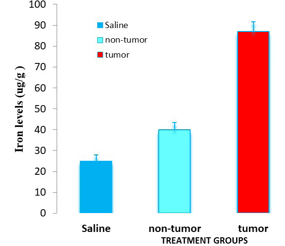
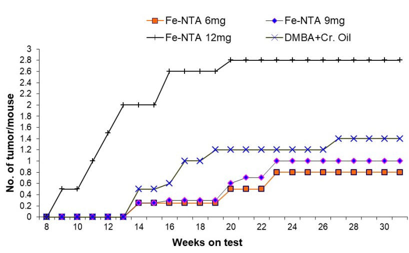
The mean ± SE of six animals is presented for each value. *p < 0.001 when compared to saline-treated normal mice; **p <0.001 when compared to saline-treated control. Effect of Fe-NTA on the Yield of Cutaneous Tumors Developed as a Result of DMBA Initiation and BPO Promotion in Mice. Figure 2 illustrates the impact of Fe-NTA on the incidence of cutaneous tumors induced by DMBA initiation and BPO promotion in various mouse groups. The group receiving DMBA, BPO, and 12 mg Fe-NTA demonstrated the first tumor in the 10th week of treatment, while no tumors were observed in the other animal groups. By week 14, the normal and other two groups receiving 6 mg or 9 mg Fe-NTA had less than 0.5 tumors per mouse, whereas, the 12 mg Fe- NTA group had roughly 2 tumors per mouse. Furthermore, tumor induction per mouse was approximately 0.8 in both control and 6 mg Fe-NTA treated animals at the 30th week, compared to 1.4 and 2.8 tumors per mouse treated with 9 and 12 mg Fe-NTA, respectively.
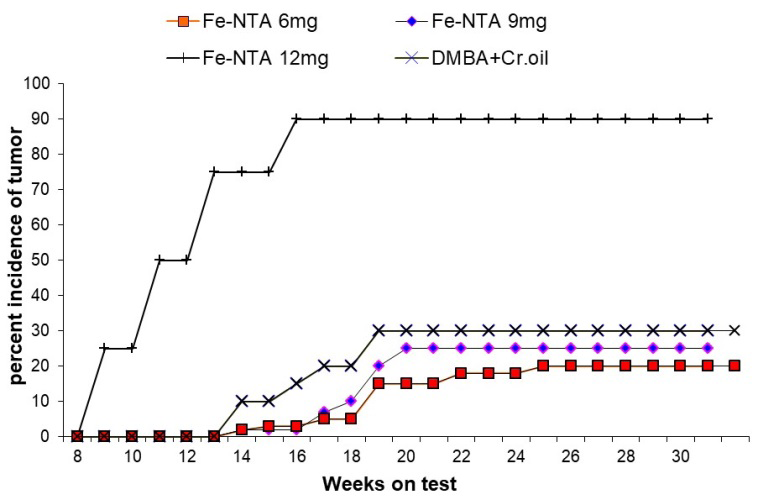
Each value represents the mean number of tumors per mouse. Effect of Fe-NTA on Percentage Incidence of Cutaneous Tumors Developed as a Result of DMBA Initiation and BPO Promotion in Mice Figure 3 shows the percentage of tumor occurrence in different mice groups. Similarly present occurrence of tumor in the 9th week among 12 mg Fe-NTA group was 25% however, at this time we did not observe any tumor sign in other three groups.
By week 14, about 75% tumor incidence in mice group treated with 12 mg Fe-NTA was noticed however, in other groups it was were less than 4%. By week 30, the percent incidences of tumor in both normal and 6 mg Fe-NTA treated animals was around 25%, compared to 30% and 90% in 9 and 12 mg Fe-NTA treated animals, respectively. As a result, the group of mice given the highest doses of Fe-NTA had the most skin tumors.
Effects of Fe-NTA on BPO-Induced Cutaneous Lipid Peroxidation in Mice: Effect of BPO on lipid peroxidation in control and Fe-NTA treated mice is shown in Table 1. Fe- NTA noticeably increases skin lipid oxidation due to BPOin comparison to other groups 6.7 ± 0.5.
| Treatment Groups | Lipid peroxidation(ng MDA/ mg protein) |
|---|---|
| Group I- Acetone | 2.5 ± 0.25 |
| Group II- BPO | 2.75 ± 0.3 |
| Group III- Fe-NTA 12mg | 3.5 ± 0.35* |
| Group IV- Fe-NTA 12mg + BPO | 7.7 ± 0.5** |
Table 1: Effect of Fe-NTA on BPO-induced cutaneous lipid peroxidation in mice.
The mean ± standard error of the mean of six animals is denoted by each value (n=6). Significance levels are *P<0.001 for comparison with normal mice treated with saline, and **P<0.05 for comparison with control mice treated with saline. Effects of Fe-NTA on BPO-induced cutaneous [3H] thymidine incorporation in mice: [3H] thymidine uptake increased in all groups in comparison to control group receiving acetone only (Table 2). Also BPO enhanced [3H] thymidine uptake in DNA of skin higher in Fe-NTA treated mice as compared to control group.
| Treatment Groups | [³H] DPM/mg Skin DNA |
|---|---|
| Group I- Acetone | 225 ± 15 |
| Group II- BPO | 240 ± 19 |
| Group III- Fe-NTA 12mg | 1480 ± 53* |
| Group IV- Fe-NTA 12mg + BPO | 2375 ± 92** |
Table 2: Effect of Fe-NTA on [3H] thymidine incorporation in cutaneous DNA in mice. Each value represents the mean ± SE of six an
Discussion
The present research offers initial proof that administering Fe-NTA topically enhances the cancer- causing capabilities of DMBA and BPO on the skin of mice. The combination of DMBA and BPO generates papilloma, which is a common model for determining the qualities of test drug. DMBA is a tumor initiator and BPO is a tumor- producing chemical that can be utilized to speed up carcinogenesis. It has been demonstrated that oxidative stress plays an important role in the tumorigenic process after DMBA and BPO treatment [17]. BPO like other known tumour promoters, inhibits junction communication, which diminishes coordination between epidermal keratinocytes [17, 18]. It induces skin irritation, dryness, erythema and contact dermatitis in humans [19].
The mechanism through which BPO promotes tumor growth is not completely known. It has been observed that, like other phorbol esters, it is activated by protein kinase C and inhibits cellular communication, both of which have been linked to tumor promoters [20]. Similarly Fe-NTA has been linked to oxidative stress and lipid peroxidation [21, 22]. Due to its geneto-toxic qualities, iron is a key factor in the development of colon cancer. In the colon cancer cell line [23], Fe-NTA functions as a free radical generator, promoting cell proliferation. Thus, BPO administration in Fe-NTA treated mice is expected to generate large amounts of free radicals and lipid peroxides, based on the current study’s findings. Furthermore, enhanced lipid peroxidation and thymidine uptake in Fe-NTA treated skin showed an increased effect of BPO tumor acceleration. In addition, lipid peroxidation and thymidine uptake are both markers of tumor promoters [24]. The findings imply that, in the presence of Fe-NTA, BPO acts as an effective tumor promoter. According to our hypothesis, iron-induced oxidative stress has a favorable reaction in accelerating BPO-mediated skin cancers.
Conclusion
The results suggest that BPO is a potent tumor promoter in the presence of Fe-NTA. This supports our hypothesis that Fe-NTA -induced oxidative stress enhances the carcinogenic effects of BPO on skin. Acknowledgment The research presented in this article was derived from a thesis (No. 4143) submitted to the Faculty of Pharmacy, Tabriz University of Medical Sciences, Tabriz, Iran, in partial fulfilment of the requirements for a Pharm D degree. The authors gratefully acknowledge the School of Pharmacy at Tabriz University of Medical Sciences for their support and provision of necessary facilities.
Conflict of Interest
The authors declare no conflict of interest.
Ethics Approval and Consent to Participate
All methods were carried out in accordance with relevant guidelines and regulations. Ethical clearance was obtained from the Regional Ethics Committee (Research Ethics Committees of Tabriz University of Medical Sciences. Ethics code: IR.TBZMED.VCR.REC.1397.191.
Funding
The authors would like to acknowledge the Vice Chancellor’s office for Research Affairs at Tabriz University of Medical Sciences for the financial support. Ethical Issues Experiments were carried out in accordance with the guide for the Care and Use of Laboratory Animals and the protocol was approved by the Committee on Animal Research of Tabriz University of Medical Sciences.
References
-
Nesslany F, Simar-Meintières S, Watzinger M, Talahari I, Marzin D (2008) Characterization of the genotoxicity of nitrilotriacetic acid. Environ Mol Mutagen 49: 439-52.
-
Mottola HA (1974) Nltrilotriacetic acid as a chelating agent: Applications, toxicology, and bio‐environmental impact. Toxicol Environ Chem 2: 99-161.
-
Okazaki Y (2022) The Role of Ferric Nitrilotriacetate in Renal Carcinogenesis and Cell Death: From Animal Models to Clinical Implications. Cancers 14: 1495.
-
Hajam YA, Rani R, Ganie SY, Sheikh TA, Javaid D (2022) Oxidative Stress in Human Pathology and Aging: Molecular Mechanisms and Perspectives. 2022 Cells 11: 552.
-
Iqbal M, Shah MS, Vun-Sang S, Okazaki Y, Okada S (2021) The therapeutic potential of curcumin in alleviating N-diethylnitrosamine and iron nitrilotriacetate induced renal cell tumours in mice via inhibition of oxidative stress: Implications for cancer chemoprevention. Biomed Pharmacother 139: 111636.
-
Khan N, Sultana S (2005) Inhibition of two stage renal carcinogenesis, oxidative damage and hyperproliferative response by Nigella sativa. Eur J Cancer Prev 14: 159-68.
-
Toyokuni S, Miyake N, Hiai H, Hagiwara M, Kawakishi S, et al. (1995) The monoclonal antibody specific for the 4‐ hydroxy‐2‐nonenal histidine adduct. FEBS letters 359: 189-191.
-
Toyokuni S, Tanaka T, Hattori Y, Nishiyama Y, Yoshida A, et al. (1997) Quantitative immunohistochemical determination of 8-hydroxy-2’-deoxyguanosine by a monoclonal antibody n45. 1: Its application to ferric nitrilotriacetate-induced renal carcinogenesis model. Lab invest 76: 365-74.
-
Iqbal M, Sharma S, Rezazadeh H, Hasan N, Abdulla M (1996) Glutathione metabolizing enzymes and oxidative stress in ferric nitrilotriacetate mediated hepatic injury. redox Report 2: 385-91.
-
Rezazadeh H, Nayebi A, Athar M (2001) Role of iron– dextran on 7, 12-dimethylbenz (a) anthracene-initiated and croton oil-promoted cutaneous tumorigenesis in normal and pregnant mice. Hum and Exp Toxicol 20: 471-76.
-
Rezazadeh H, Athar M (1998) Effect of iron overload on the benzoyl peroxide-mediated tumor promotion in mouse skin. Cancer Letters 1998; 126: 135-42.
-
Awai M, Narasaki M, Yamanoi, Seno S (1979) Induction of diabetes in animals by parenteral administration of ferric nitrilotriacetate. A model of experimental hemochromatosis. Am J Pathol 95: 663.
-
Connell JF, Klein-Szanto AJ, DiGiovanni DM, Fries JAW, Slaga TJ (1986) Enhanced malignant progression of mouse skin tumors by the free-radical generator benzoyl peroxide. Cancer Res 46: 2863-65.
-
Wright JR, Colby HD, Miles PR (1981) Cytosolic factors which affect microsomal lipid peroxidation in lung and liver. Arch Biochem Biophys 206: 296-304.
-
Smart RC, Huang MT, Conney AH (1986) Sn- l, 2-diacylglycerols mimic the effects of 12-0-tetradecanoylphorbol-13-acetate in vivo by inducing biochemical changes associated with tumor promotion in mouse epidermis. Carcinogenesis 7: 1865- 70.
-
Giles KW, Myers A (1965) An improved diphenylamine method for the estimation of deoxyribonucleic acid. Nature 206: 93.
-
Slaga TJ, Klein-Szanto AJP, Triplet LL (1981) Skin tumor- promoting activity of benzoyl peroxide, a widely used free radical-generating compound. Science 213: 1023- 1025.
-
Hartman TG, Rosen JD (1983) Inhibition of metabolic cooperation by cigarette smoke condensate and its fractions in V-79 Chinese hamster lung fibroblasts. Proc Natl Acad Sci of the USA 80: 5305-5309.
-
Kawashima M, Nagare T, Doi M (2017) Clinical efficacy and safety of benzoyl peroxide for acne vulgaris: comparison between Japanese and Western patients. J Dermatol 44: 1212-1218.
-
Jansen LA, Mesnil M, Jongen WM (1996) Inhibition of gap junctional intercellular communication and delocalization of the cell adhesion molecule E-cadherin by tumor promoters. Carcinogenesis 17: 1527-1531.
-
Ansar S, Iqbal M, AlJameil N (2014) Diallyl sulphide, a component of garlic, abrogates ferric nitrilotriacetate- induced oxidative stress and renal damage in rats. Human and Experimental Toxicology 33: 1209-1216.
-
Ansar S, Iqbal M (2016) Amelioration of ferric nitrilotriacetate-induced hepatotoxicity in Wistar rats by diallylsulfide. Hum Exp Toxicol 35: 259-266.
-
Knöbel Y, Glei M, Osswald K, Pool-Zobel BL (2006) Ferric iron increases ROS formation, modulates cell growth and enhances genotoxic damage by 4-hydroxynonenal in human colon tumor cells. Toxicol In Vitro 20: 793-800.
-
Gimenez-Conti I, Viaje A, Chesner J, Conti C, Slaga TJ (1991) Induction of short-term markers of tumor promotion by organic peroxides. Carcinogenesis 12: 563-569.
- Pattern of Gonadal Hormones in Oral Testosterone-Supplimented Male Wistar Rats with Diabetes-Induced Hypogonadism
- Re-Evaluation of the Genotoxicity of Currently Used Food Dyes in Mouse Multiple Organs Via Continuous Administration by Drinking Using the Comet Assay
- Pharmacogenetics of Type 2 Diabetes Mellitus: Linking Genetic Variability to Drug Efficacy and its Cardiovascular Outcomes
- Exploratory Proteomic Profiling of SARS-CoV-2 Infected THP-1 Macrophages Reveals Alterations in Inflammatory Response and Cellular Metabolism
- Study of Genotoxicity of Hepatocarcinogens in Multiple Organs in Mice by Feeding and Drinking Using the Comet Assay
- Spirulina Polypeptides Inhibit the Growth of Human Lung Tumor (H460) Cells