Comparative Investigation of In Vitro Induction of DNA Damage and Micronuclei by Pro-Mutagens in Human-Derived Hepatoma HepG2 Cells
It is important to know whether the DNA initial lesions are repaired or derive into chromosome aberration, gene mutation and/or cell death to discuss chemical carcinogenicity. To know whether DNA damage detected by the comet assay is repaired or fixed, the results of micronucleus (MN) test was compared to those of the comet assay in HepG2 cells. HepG2 cells were exposed to 38 pro-mutagens for 4 h followed by the cultivation for 24 h with cytochalasin B and the results were compared to those of the comet assay shown in our previous study where HepG2 cells were exposed to same 38 pro-mutagens for 4 h followed by immediate sampling. Among 38 pro-mutagens studied, 35 were positive in the comet assay and 22 were positive in the MN test. Since for 7 pro-mutagens, the lowest comet-positive concentration was higher than MN-positive concentration range, it is difficult to declare simply that the detecting ability of the comet assay is superior to that of the MN test. Comet assay-detected DNA damages that were induced by aromatic nitro compounds, azo dyes, heterocyclic amines, and aromatic hydrocarbons having bay structure, and aromatic amines with condensed multiple ring tended to result in cytotoxicity rather than MNs. It was considered that one important factor to decide the fate to produce MNs of comet-detected DNA damages might be whether detected DNA damages are bulky base adducts at non-cytotoxic concentration range and that chemical structures of pro-mutagens are important factor to decide whether induced base adducts are bulky.
Abbreviations
DBDs: Double Strand Breaks; SSBs: Single Strand Breaks; BNC: Bi-Nuclei Cells.
Introduction
To discuss chemical carcinogenicity, it is important to know whether DNA initial lesions are repaired or produce chromosome aberration, gene mutation and/or cell death.
Although DNA double strand breaks (DDBs) are the principal lesions in the process to induce chromosome aberration- formation [1], most chemical mutagens cannot induce DDBs directly but initial lesions such as base adducts and AP- sites. Such initial lesions produce DNA single strand breaks (SSBs) and DDBs during repair or DNA synthesis and result in chromosome aberrations [1]. Many methods have been used to identify genotoxic substances. They include methods to detect DNA damages, chromosome aberrations, and gene mutations in both in vitro and in vivo. Although the comet assay can detect DNA initial lesions such as base adducts, AP sites, and DSBs [2, 3], it cannot provide any information about the fate of DNA lesions. Therefore, comparative study of the comet assay and MN test would be able to provide good information about the fate of initial DNA lesions. In this study, we focused on whether comet assay-detected DNA lesions induced by pro-mutagens can derive into MNs and/ or cytotoxicity. We have previously shown that various pro- mutagens having different kinds of functional groups can induce DNA initial lesions in HepG2 cells and that HepG2 cells could be usefully used to detect the genotoxicity of pro- mutagens for the purpose of the evaluation of the genotoxic risk for humans [4]. HepG2 cells have been isolated 30 years ago from a hepatoblastoma of an 11-year-old boy from Argentina [5]. HepG2 cells retain many of the morphological characteristics of liver parenchymal cells and contain several enzymes responsible for the activation of various pro- mutagens [6]. Therefore, if the results of the comet assay were compared to those of the MN test under the identical exposure condition, it would be possible to know the fate of DNA initial lesions detected by the comet assay in HepG2 cells. The aim of this study was to analyse the fate of DNA initial lesions by adding the results of the MN test to those of the comet assay previously shown. In the comet assay, relative survivals immediately after the exposure to pro- mutagens were used as the parameter of cytotoxicity. In the MN test, on the other hand, the frequency bi-nuclei cells can be used as the indicator of cytotoxicity, since cells that had divided once after chemical treatment can be distinguished from those that had not divided by blocking the cytokinesis [7].
Materials and methods
Chemicals, cells and medium
Table 1 shows the pro-mutagens tested, their abbreviations and CAS numbers.
| Promutagens | Abbreviation | CAS number | Sourcea | Vehicleb |
|---|---|---|---|---|
| Aromatic hydrocarbons without bay region | ||||
| Benzene | 71-43-2 | W | D | |
| Pyrene | 129-00-0 | T | D | |
| Aromatic hydrocarbons with bay region | ||||
| Benz[a]anthrathene | B[a]A | 56-55-3 | T | D |
| Benzo[a]pyrene | B[a]P | 50-32-8 | S | D |
| Phenanthrene | 85-01-8 | T | D | |
| Pyrene | 129-00-0 | T | D | |
| One ring aromatic amines | ||||
| Aniline | 62-53-3 | W | D | |
| 2,4-Diaminotoluene | 2,4DAT | 95-80-7 | W | D |
| Phenacetin | 62-44-2 | W | D | |
| Condensed two rings aromatic amines | ||||
| 1-Naphthylamine | 1NA | 134-32-7 | T | D |
| 2-Naphthylamine | 2NA | 91-59-8 | * | D |
| Condensed multiple rings aromatic amines | ||||
| 2-Aminoanthracene | 2AA | 613-13-8 | W | D |
| 1-Aminopyrene | 1AP | 1606-67-3 | W | D |
| Benzidine derivatives | ||||
| Benzidine | 92-87-5 | * | D | |
| 3,3’-Dichlorobenzidine | DCB | 91-94-1 | S | D |
| o-Tolidine | 119-93-7 | T | D | |
| Heterocyclic amincs | ||||
| Trp-P-1 acetate | 68808-54-8 | W | S | |
| Trp-P-2 acetate | 62450-10-3 | W | S | |
| AαC | 26148-68-5 | W | D | |
| IQ | 76180-96-6 | W | D | |
| MeIQ | 77094-11-2 | W | D | |
| PhIP hydrochloride | 105650-23-5 | W | S | |
| Linear dialkyl N-nitrosamines | ||||
| N-Nitrosodimethylamine | DMN | 62-75-9 | W | S |
| N-Nitrosoethylmethylamine | EMN | 10595-95-6 | W | S |
| N-Nitrosodiethyamine | DEN | 55-18-5 | W | S |
| N-Nitrosodiethanolamine | DEolN | 1116-54-7 | S | S |
| N-Nitrosodipropylamine | DPN | 621-64-7 | N | D |
| N-Nitrosodibutylamine | DBN | 924-16-3 | S | D |
| Cyclic alkyl N-nitrosamines | ||||
| N-Nitrosomorpholine | NMOR | 59-89-2 | S | S |
| N-Nitrosopiperidine | NPIP | 100-75-4 | Ka | S |
| N-Nitrosopyrrolidine | NPYR | 930-55-2 | Ka | D |
| Azo dyes | ||||
| Azobenzene | 103-33-3 | T | D | |
| p-Aminoazobenzene | pAAB | 60-09-3 | W | D |
| p-Dimethylaminoazobenzene | DAB | 60-11-7 | W | D |
| Nitro compounds | ||||
| 1-Nitronaphtharene | 1NN | 86-57-7 | T | D |
| 1-Nitropyrene | 1NP | 5522-43-0 | T | D |
| Vinyl compounds | ||||
| Acrylamide | 79-06-1 | W | D | |
| Styrene | 100-42-5 | W | D | |
| Others | ||||
| Aflatoxin B1 | AFB | 1162-65-8 | W | S |
| 1,2-Dimethylhydrazine dihydrochloride | DMH | 540-73-8 | W | S |
Table 1: ** Pro-mutagens tested in this study.
aK: Kanto Chemical Co., Inc., Tokyo (Japan); Ka: Katayama Chemical; S: Sigma Chemical Co., St. Louis, MO (U.S.A.);T: Tokyo Kasei Kogyo, Tokyo (Japan); W: Wako Pure Chemical Co (Osaka); *2-Naphthylamine was synthethized from 2-naphththol and ammonium sulfite in Laboratory of Genotoxicity, National Institute of Technology, Hachinohe College. Benzidine was synthethized from hydrazobenzene in Laboratory of Genotoxicity, National Institute of Technology, Hachinohe College. bD, DMSO; S, Saline. Table 1: Pro-mutagens tested in this study.
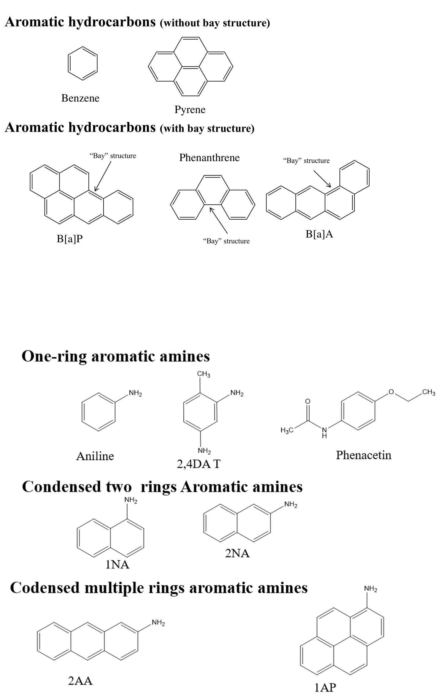
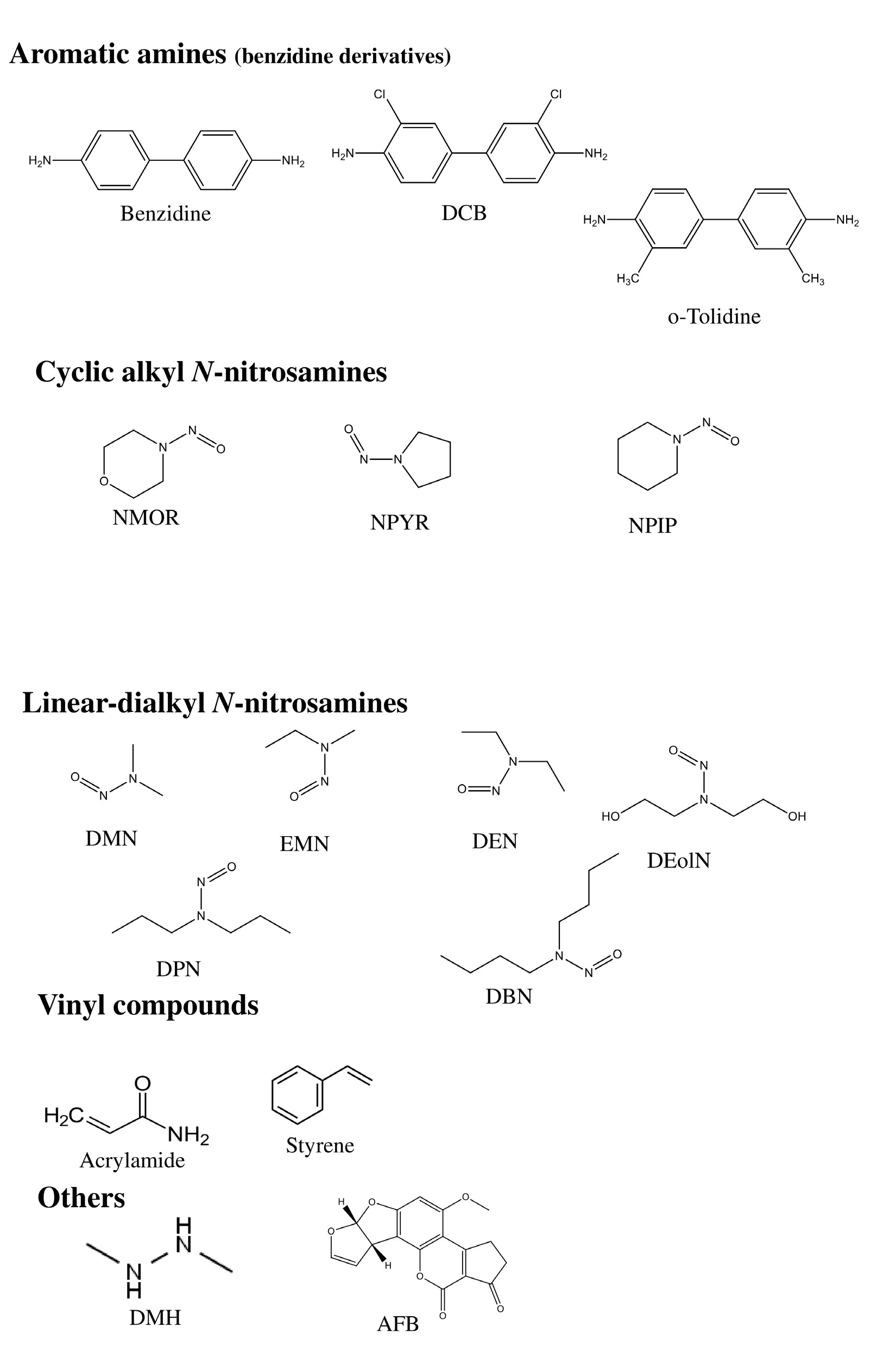
Studied pro-mutagens divided into 8 categories based on their structures ; i.e. (1) aromatic hydrocarbon, (2) aromatic amine, (3) alkyl N-nitrosamine, (4) aromatic nitro compound, (5) azo dyes, (6) heterocyclic amine, (7) vinyl compound, and (8) other. Aromatic hydrocarbon includes 2 sub-categories; i.e, aromatic hydrocarbon with and without bay structure. Bay structure means a recess formed by three condensed rings (Figure 1). Aromatic amine includes 4 sub-categories;
i.e, one ring aromatic amine, condensed two rings aromatic amine, condensed multiple rings aromatic amine aromatic amine, and benzidine derivative. Alkyl N-nitrosamine includes 2 sub-categories; i.e, linear-dialkyl N-nitrosamines and cyclic-alkyl N-nitrosamines. Their structures are shown in Figure 1.
![Figure 3: Pro-mutagens showed positive responses in MN test and comet assay in HepG2 cells. The data of comet assay are cited from previous results [4].](/fulltextimages/13700/fig_3.jpeg)
Human hepatoma (HepG2) cells, obtained from Riken Cell Bank, Tokyo, were grown in 1:1 mixture medium of Dulbecco MEM and Ham’s F12 supplemented with 10% fetal bovine serum (HyClone Laboratories, Inc., U.S.A.). All media were purchased from Nissui Pharmaceutical Inc., Tokyo. Cells were grown at 37oC in a humidified atmosphere containing 5% CO2.
MN Test
To compare the results with those of the comet assay, cells were treated under the same conditions as in the comet assay [4]: i.e., HepG2 cells were exposed for 4 h to the different test compounds or to the vehicle control. Although cells were sampled immediately after the treatment in the comet assay, in the MN test the cells were washed with Hanks’ BSS at the end of a 4 h treatment period, followed by the cultivation for 24 h in the medium containing cytochalasin B at 3µg/mL [8]. After the cultivation, collected cells were suspended in 0.075M KCl hypotonic solution for 15 min. Then, cells were re-collected, fixed by 10% neutral formalin, and were re-suspended in 0.075M KCl solution containing 10% (v/v) DMSO followed by the freeze and store at –80oC until the observation. Cells were stained by a method modified from that used in an MN test for peripheral blood cells reported by Hayashi, et al. [9]. Briefly, immediately before the analysis, frozen cell suspension was dissolved and concentrated to the volume of 50 µL, and then 20 µL of cell suspensions were put onto slide glass coated with acridine orange and mounted with 24 x 48 mm cover slips. Bi- nuclei cells with MNs (MNBNC) at 1000 bi-nuclei cells (BNC) and BNC at 1000 cells were scored with the aid of fluorescence microscope (Olympus at 600x magnification) equipped with G filter. The incidence of MNBNC was analyzed statistically by ANOVA and the Dunnett test. The relative cytotoxicity to control was obtained as the frequency of BNC at 1000 cells.
Results
Because the purpose of this study was to compare the results for comet assay and MN test under the same exposure conditions, present results of MN test [MNBNC (‰) and relative BNC (%)] were shown overlaid on those of comet assay [tail length and relative survival (%)] cited from our previous study [4]. Figures 2 A-2G shows the concentration- response curves of the test compounds in the MN assays. Relative BNC less than 50% was considered to show remarkable cytotoxicity. While relative survivals in the comet assay show cell survival immediately after the exposure to pro-mutagens, relative BNC in the MN test show cytotoxicity after the cultivation for 24 h following to the exposure to pro- mutagens.
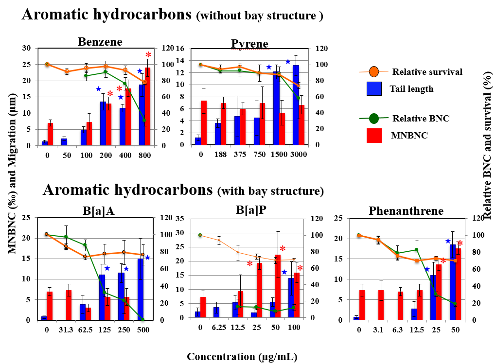
Figure 2A: The results MN test for aromatic hydrocarbons in HepG2 cells overlaid on the results of comet assay (tail length and relative survival). Each graph shows Mean ± SD for 3 independent experiments. Significant difference from untreated control: p<0.05; *for MNBNC in MN test :★for tail length in the comet assay. The results of the comet assay were cited from previous results [4].
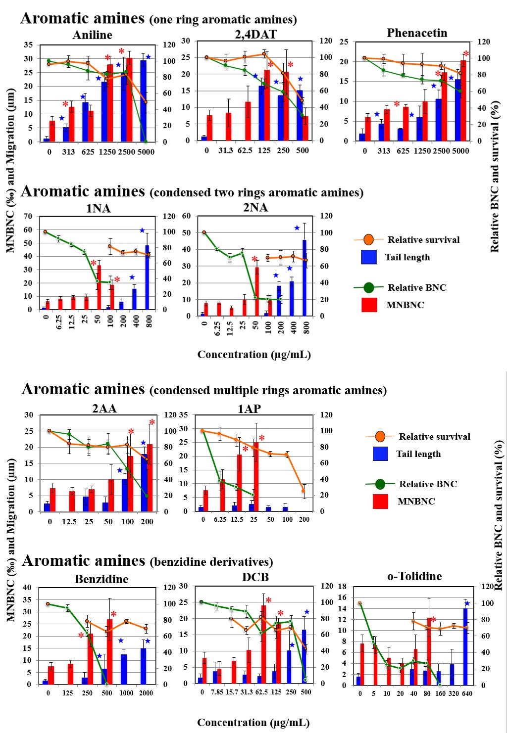
Figure 2B: The results MN test for aromatic amines in HepG2 cells overlaid on the results of comet assay (tail length and relative survival). Each graph shows Mean ± SD for 3 independent experiments. Significant difference from untreated control: p<0.05; *for MNBNC in MN test :★for tail length in the comet assay. The results of the comet assay were cited from previous results [4].
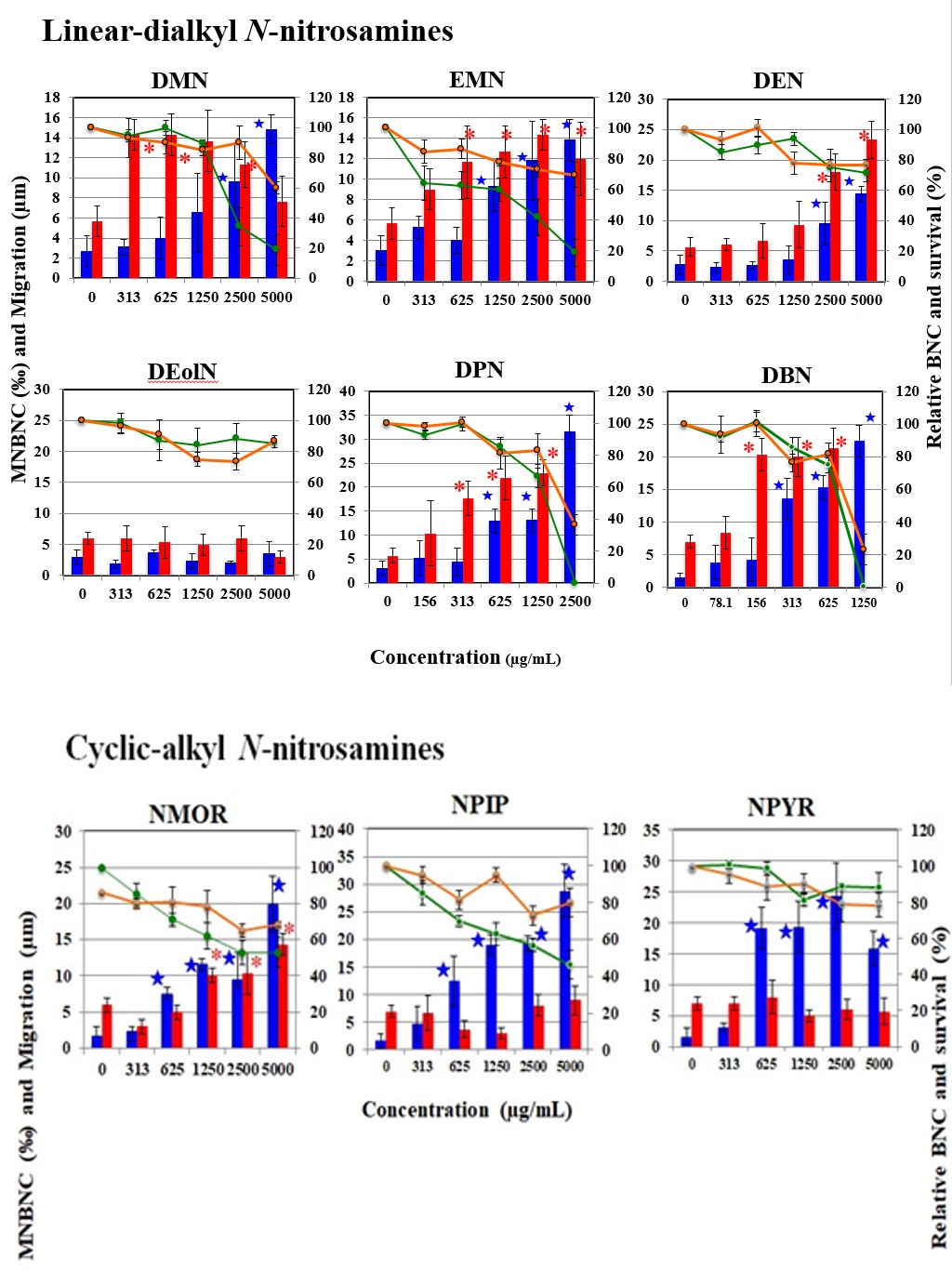
Figure 2C: The results MN test for linear-alkyl N-nitrosamines in HepG2 cells overlaid on the results of comet assay (tail length and relative survival). Each graph shows Mean ± SD for 3 independent experiments. Significant difference from untreated control: p<0.05; *for MNBNC in MN test, :★for tail length in the comet assay. The results of the comet assay were cited from previous result.
Figure 2D: The results MN test for cyclic-alkyl N-nitrosamines in HepG2 cells overlaid on the results of comet assay (tail length and relative survival). Each graph shows Mean ± SD for 3 independent experiments. Significant difference from untreated control: p<0.05; *for MNBNC in MN test, :★for tail length in the comet assay.The results of the comet assay were cited from previous results [4].
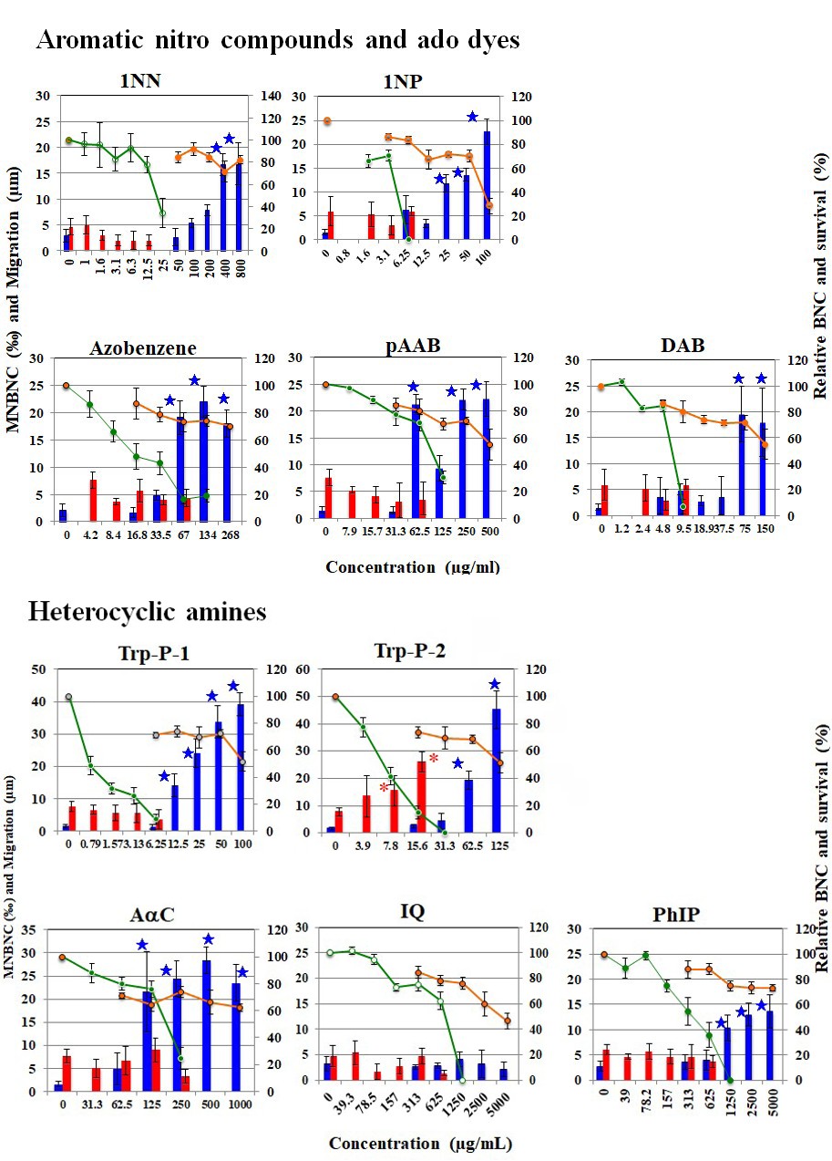
Figure 2E: The results MN test for aromatic nitro compounds and azo dyes in HepG2 cells overlaid on the results of comet assay (tail length and relative survival). Each graph shows Mean ± SD for 3 independent experiments. Significant difference from untreated control: p<0.05; *for MNBNC in MN test, :★for tail length in the comet assay. The results of the comet assay were cited from previous results [4].
Figure 2F:The results MN test for heterocyclic amines in HepG2 cells overlaid on the results of comet assay (tail length and relative survival). Each graph shows Mean ± SD for 3 independent experiments. Significant difference from untreated control: p<0.05; *for MNBNC in MN test, :★for tail length in the comet assay. The results of the comet assay were cited from previous results [4].
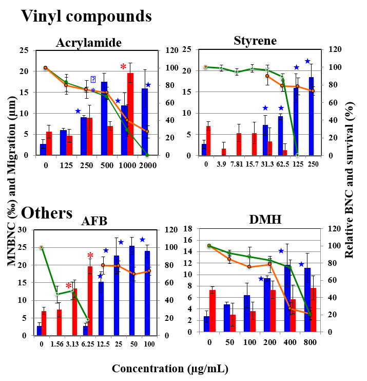
Figure 2G: The results MN test for vinyl compounds and other compounds in HepG2 cells overlaid on the results of comet assay (tail length and relative survival). Each graph shows Mean ± SD for 3 independent experiments. Significant difference from untreated control: p<0.05; *for MNBNC in MN test, :★for tail length in the comet assay. The results of the comet assay were cited from previous results [4].
Aromatic Hydrocarbons
Benzene and phenanthrene showed positive response in both comet assay and MN test at almost identical dose range (200, 400 and 800 µg/mL for benzene and 25 and 50 µg/mL for phenanthrene). Although B[a]P at ≥12.5 µg/ mL decreased relative BNC to <20%, it increased MNBNC at ≥25 µg/mL and increased tail length at 100 µg/mL. Pyrene at >1500 µg/mL increased tail length but not MNBNC. Although B[a]A at ≥125 µg/mL decreased BNÇ to < 40%, it increased tail length but not MNBNC.
Aromatic Amines
Aniline, 2,4DAT, 2AA, and phenacetin showed positive response in both comet assay and MN test at almost identical dose range. Although 1AP at >12.5 µg/mL decreased relative BNC to <40%, it increased MNBNC but not tail length. 1NA, 2NA, and three benzidine compounds (benzidine, DCB, and o-tolidine) increased tail length and MNBNC. The comet- positive concentration range did not overlap with and was higher than MN-positive concentration range and the relative BNC decreased remarkably at the comet-positive concentration range.
Linear- dialkyl N-nitrosamines
DMN, EMN, DEN, DPN, and DBN showed positive response in both comet assay and MN test at almost identical dose range. DEolN did not show positive responses neither comet assay nor MN test at up to 5000 µg/mL.
Cyclic-alkyl N-nitrosamines
NMOR showed positive response in both comet assay and MN test at almost identical dose range. NPIP and NPYR showed positive response in the comet assay but not in MN test (they did not increase MNBNC at up to 5000 µg/mL).
Aromatic Nitro Compounds and Azo Dyes
In this study, two aromatic nitro compounds (1NN and 1NP) and three azo dyes (azobenzene, pAAB, and DAB) were tested. They yielded positive responses in the comet assay but not in the MN test. They decreased relative BNC to <40% at the comet-positive concentration ranges (≥400 µg/mL for 1NN, ≥25 µg/ml for 1NP, ≥67 µg/mL for azobenzene, ≥62.5 µg/mL for pAAB, and ≥75 µg/mL for DAB).
Heterocyclic Amines
Although Trp-P-2 decreased relative BNC to <40% at 7.8 and 15 µg/mL, it increased MNBNC at 7.8 and 15 µg/mL and tail length at 62.5 and 125 µg/mL. The comet-positive concentration range did not overlap with and was higher than MN-positive concentration range and the relative BNC decreased remarkably at the comet-positive concentration range. Although Trp-P-1 and AαC increased tail length at ≥12.5 µg/mL and ≥125 µg/mL, respectively, they did not increase MNBNC. PhIP at ≥1250 µg/mL increased tail length but not MNBNC. IQ did not induce MNBNC and tail length at up to 5000 µg/mL.
Vinyl Compounds and Others
Acrylamide increased tail length at 250 - 2000 µg/mL and MNBNC at 1000 µg/mL. Styrene induced tail length but not MNBNC at ≥31.3 µg/mL. AFB increased tail length at 3.13 and 6.25 µg/mL and it increased MNBNC at ≥12.5 µg/mL. The comet-positive concentration range did not overlap with and was higher than MN-positive concentration range and the relative BNC decreased remarkably at the comet-positive concentration range. DMH at ≥200 µg/mL induced tail length but it did not increased MNBNC.
Discussion
When the power to detect the genotoxicity of pro- mutagens is compared between the comet assay and MN test under identical treatment period, 35 pro-mutagens were positive in the comet assay and 22 were positive in the MN test. Out of 35 comet-positive pro-mutagens, 21 were positive in MN test (Figure 3).
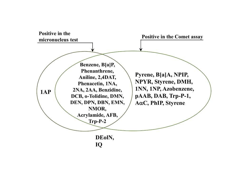
Therefore, the detecting ability of the former seems to be superior to that of the latter. Among 21 pro-mutagens that were positive in both assays, however, for 7 pro-mutagens (B[a]P, 1NA, 2NA, DCB, Tolidine, Trp-P-2, and AFB), the lowest comet-positive concentration was higher than MN- positive concentration range. The comet assay detects DNA lesions immediately after the end of the exposure to mutagens, whereas the MN test detects structure chromosome aberrations as MNs after a 24-hour recovery time. Even if relative survivals do not decrease immediately after the end of the exposure to mutagens, some survived cells having DNA damage could be considered not to enter to next cell cycle during 24-hour recovery time. Therefore, it is difficult to declare simply that the power of the comet assay to detect genotoxicity is superior to that of the MN test. The comet assay detects primary DNA damage, while the MN test detects structural chromosome aberrations that are derived from primary lesions in the S phase and/or numerical chromosome aberrations due to aneugenic effects in the M phase [1, 7]. Comet assay-detected damage would include damage that derive into structural chromosome aberrations and that does not. The latter might include DNA lesions that is repaired before DNA replication and pull a trigger of cell death and/or prolong cell cycle duration. When the results of the MN test are compared with those of the comet assay, studied pro-mutagens were divided into seven categories (Figure 4).
(1) 14 Pro-mutagens (Benzene, Aniline, 2,4DAT, Phenanthrene, Phenacetin, Benzidine, EMN, 2AA, DEN, DPN, DMN, Acrylamide, DBN, and NMOR) were positive in the comet assay and the MN test at almost identical non-cytotoxic concentration range where remarkable decreases in relative BNC were not observed. Their induced comet assay-detected DNA damages were considered to produce MNs rather than cytotoxicity. (2) One Pro-mutagen (B[a]P) was positive in the comet assay and the MN test at almost identical cytotoxic concentration where remarkable decrease in relative BNC was observed. Its induced DNA damage detected by the comet assay that did not cause cell death and was considered to cause MNs. (3) 8 Pro-mutagens (Pyrene, NPIP, NPYR, DMH, 1NP, Azobenzene, pAAB, and DAB) show neither MN- induction nor cytotoxicity at the concentration range where the comet assay was positive.
It is considered that their induced comet assay-detected DNA damages are repaired or that the level of DNA damage is too small to produce MNs that can be observed by microscope. (4) 6 Pro-mutagens (1NA, 2NA, Trp-P-2, AFB, DCB, and o-Tolidine) decreased relative BNC remarkably at the concentration range where the comet assay was positive but induced MNs at the concentrations range lower than that where the comet assay was positive. It would be considered that their induced comet assay-detected DNA damage cause cytotoxicity and that comet assay undetectable DNA damage produce MNs. (5) 6 Pro-mutagens (B[a]A,1NN, Styrene, Trp-P-1, AαC, and PhIP) decreased relative BNC remarkably at the concentration range where the comet assay was positive but did not induce MNs at any concentration range. Comet assay detected DNA damages were considered to produce cell death but not MNs. (6) Only 1 (1AP) was positive in the MN test but not in the comet assay. (7) DEolN and IQ were negative in the comet assay and MN test.
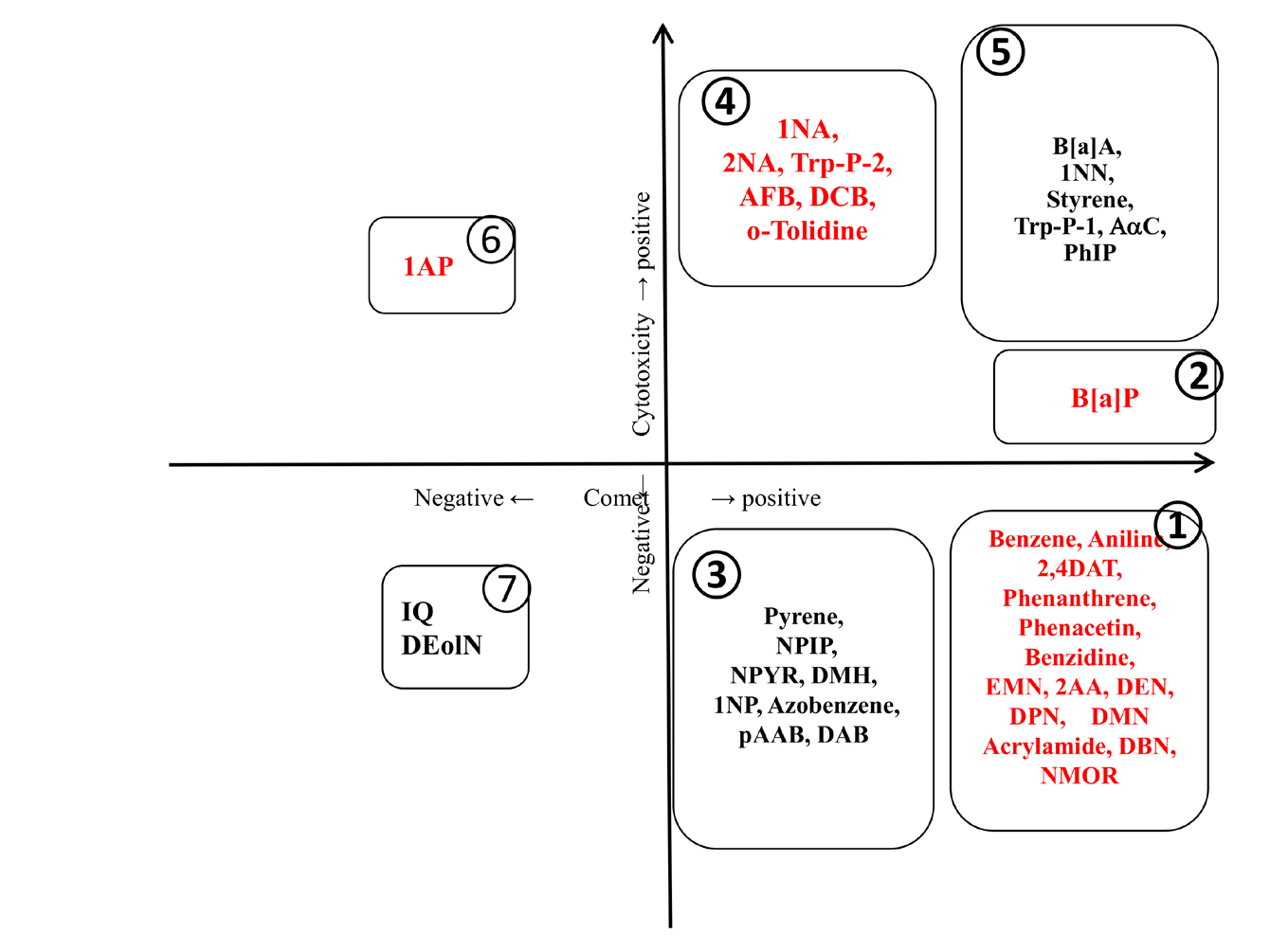
Based on above categories, aromatic nitro compounds, azo dyes, and heterocyclic amines excluding IQ decreased relative BNC remarkably at the concentration range where the comet assay was positive (category 6), suggesting that their induced comet assay-detected DNA damage result in cytotoxicity rather than MNs. In studied aromatic hydrocarbons, benzene was positive in the comet assay and MN test at the concentration range not showing cytotoxicity (category 1). Pyrene was positive in the comet assay but not MN test at the concentration range not showing cytotoxicity (category 3). Benzene and pyrene do not have bay structure, on the other hand, phenanthrene (category 2), B[a]P (category 2), and B[a]A (category 5) that have bay structure were positive in the comet assay and/or MN test at the concentration range showing cytotoxicity. Considering that the bay structure of aromatic hydrocarbons is bio- transformed to electrophilic diol epoxides that can react with DNA [10], higher reactivity of their metabolites may responsible for their greater cytotoxicity.
In aromatic amines, the fate of DNA damage detected in the comet assay might depends on the number of aromatic ring. One-ring aromatic amines (aniline, 2,4DAT, and phenacetin) induced DNA damage and MNs at almost identical non-cytotoxic concentration range (category 1). Aromatic amines having condensed 2 aromatic rings (1NA and 2NA), condensed multiple aromatic rings (1AA and 2AP), and benzidine derivatives, decreased relative BNC greatly at the concentration range where the comet assay but not MN test was positive (category 3), suggesting that their induced comet assay-detected DNA damage tend to produce cytotoxicity rather than MNs. They induced MNs at the concentration range where comet assay was negative, suggesting that DNA damage whose level is too small to be detected by the comet assay could produce MNs.
Genotoxicity and cytotoxicity of N-nitrosamines largely depends on the structure of alkyl moiety. While comet- detected DNA damage induced by N-nitrosamines with cyclic moiety might not produce MNs, comet-detected DNA damage induced by N-nitrosamine with linear alkylating moiety might produce MNs. The size of linear alkylating moiety affected genotoxicity and cytotoxicity: i.e., DPN and DBN were more cytotoxic than DMN, EMN, and DEN. In the comet assay, the lowest positive concentrations of DPN (625 µg/mL) and DBN (313 µg/mL) were lower than those of DEN
and DMN (2500 µg/mL). In the MN test, the lowest positive concentrations of DPN and DBN (313 µg/mL for DPN, 156 µg/mL for DBN) were lower than those of DEN (2500 µg/ mL) and DMN (313 µg/mL). Therefore, our results suggested that chemical structures of pro-mutagens tended to affect the fate of DNA damage detected by the comet assay. It is known that a number of bulky base adducts produced by benzene metabolites can inhibit topoisomerase II activity, which may represent a potential mechanism for clastogenic effects of benzene [11, 12]. Also, we have already shown that bulky base adducts causing disturbances to the helical DNA structure result in clastogenicity rather than mutagenicity [13]. Therefore, pro-mutagen metabolites that produce bulky base adducts causing disturbances to the helical DNA structure could be considered to produce structural chromosome aberrations that are detected as MNs at non- cytotoxic concentration range. Based on those discussion, one important factor to decide the fate to produce MNs of comet-detected DNA damages might be whether detected DNA damages are bulky base adducts that cause disturbances to the helical DNA structure and their induction level at non- cytotoxic concentration range is high enough to produce DDBs during next S phase and retain until next M phase. Even if comet-detected DNA damages are non-bulky adducts, for example, methylated bases, when their induction level is high enough to produce DDBs during next S phase and retain until next M phase, they could produce MNs. As we have shown [14], chemical structures of pro-mutagens are important factor to decide whether induced base adducts are bulky to cause disturbances to the helical DNA structure. To discuss the toxicological meaning of the results of the comet assay, it is important to clarify the fate of comet assay-detected DNA damage. As shown in this study, since the endpoints of comet assay and MN test are different, comparative investigation by both systems would provide good information for genotoxicity. When the results of the comet assay and the MN test were compared at the same concentration range, comet assay detected DNA damages induced by one-ring aromatic amines and long-chain alkylnitrosamines tended to produce MNs, whereas those induced by condensed aromatic hydrocarbons, cyclic alkylnitrosamines, and azo compounds tended not to produce MNs. From the above, it was considered that the fate of the initial DNA damage in HepG2 cells depends on the structures of pro-mutagens.
Acknowledgements
This research was conducted in Advanced Material and Biological Engineering Course, National Institute of Technology, Hachinohe College as part of a graduation research project by Saeko Okutani under the research guidance of course staffs based on allocation of school educational expenses. The authors acknowledge the Advanced Material and Biological Engineering Course, National Institute of Technology, Hachinohe College.
References
-
Obe G, Pfeiffer P, Savage JRK, Johannes C, Goedecke W, et al. (2002) Chromosomal aberrations: formation, identification and distribution. Mutation Res 504(1-2): 17-36.
-
Singh, NP, McCoy MT, Tice RR, Schneider EL (1988) A simple technique for quantitation of low levels of DNA damage in individual cells. Exp Cell Res 175(1): 184-191.
-
Fairbairn DW, Olive PL, Neill KL (1995) The comet assay: a comprehensive review. Mutation Res 339: 37-59.
-
Kawaguchi S, Okutani S, Yamamoto A, Nakamura T, Sasaki YF (2024) Comparative Investigation of In Vitro Genotoxicity of Pro-Mutagens Using Human-Derived Hepatoma HepG2 Cells with Human and Rat Liver S9. ACT 9(3): ACT16000314.
-
Aden DP, Fogel A, Plotkin S, Damjanov I, Knowles BB (1979) Controlled synthesis of HBs Ag in a differentiated human liver carcinoma-derived cell line. Nature 282: 615-616.
-
Knasmuller S, Parzefall W, Sanyal R, Ecker S, Schwab C, et al. (1998) Use of metabolically competent human hepatoma cells for the detection of mutagens and antimutagens. Mutation Res 402(1-2): 185-202.
-
Fenech M, Morley AA (1985) Measurement of micronuclei in lymphocytes. Mutation Res 147: 29-36.
-
Falck G, Catalan J, Norppa H (1997) Influence of culture time on the frequency and contents of human lymphocyte micronuclei with and without cytochalasin. Mutation Res 392(1-2): 71-79.
-
Hayashi M, Morita T, Kodama Y, Sofuni T, Ishidate M Jr (1990) The micronucleus assay with mouse peripheral blood reticulocytes using acridine orange-coated slides. Mutation Res 245: 245-249.
-
Jerina DM, Sayer JM, Agarwal SK, Yagi H, Levin W, et al. (1986) Reactivity and tumorigenicity of bay-region diol epoxides derived from polycyclic aromatic hydrocarbons. Adv Exp Med Biol 197: 11-30.
-
Hutt AM, Kalf GF (1996) Inhibition of human DNA topoisomerase II by hydroquinone and p-benzoquinone, reactive metabolites of benzene. Environmental Health Perspectives 104(6): 1265-1269.
-
Hang B (2010) Formation and repair of tobacco carcinogen-derived bulky DNA adducts. J Nucleic Acids. J
- Pattern of Gonadal Hormones in Oral Testosterone-Supplimented Male Wistar Rats with Diabetes-Induced Hypogonadism
- Re-Evaluation of the Genotoxicity of Currently Used Food Dyes in Mouse Multiple Organs Via Continuous Administration by Drinking Using the Comet Assay
- Pharmacogenetics of Type 2 Diabetes Mellitus: Linking Genetic Variability to Drug Efficacy and its Cardiovascular Outcomes
- Exploratory Proteomic Profiling of SARS-CoV-2 Infected THP-1 Macrophages Reveals Alterations in Inflammatory Response and Cellular Metabolism
- Study of Genotoxicity of Hepatocarcinogens in Multiple Organs in Mice by Feeding and Drinking Using the Comet Assay
- Spirulina Polypeptides Inhibit the Growth of Human Lung Tumor (H460) Cells