Improvement of Vitamin D3 Metabolites Level after Oral Administration of the Biofield Energy Treated Proprietary Test Formulation in Vitamin D3 Deficiency Diet (VDD) Induced Rat Model
The present experiment was evaluated to study of the effect of Consciousness Energy Healing Treatment (the Trivedi Effect®) on novel proprietary test formulation in male Sprague Dawley (SD) rats, fed with vitamin D3 deficiency diet (VDD) for the estimation of vitamin D3 metabolites in kidney tissue. A novel proprietary test formulation was formulated with a few minerals (magnesium, zinc, copper, calcium, selenium, and iron), vitamins (ascorbic acid, pyridoxine HCl, alpha tocopherol, cyanocobalamin, and cholecalciferol), Panax ginseng extract, β-carotene, and cannabidiol isolate. The novel test formulation was divided into two parts; one part was defined as the untreated test formulation, while the other part of the test formulation and the animals received Biofield Energy Healing Treatment by a renowned Biofield Energy Healer, Mr. Mahendra Kumar Trivedi. 25-OH vitamin D3 level was significantly increased in the Biofield Energy Treated test formulation group (G5), Biofield Energy Treatment per se (G6), and 15-days pre-treatment of the Biofield Energy Treated test formulation (G7) groups by 21%, 22.9%, and 11.5%, respectively as compared with the disease control group (G2). Additionally, the level of 1,25-(OH)2D3 was increased by 55% in the G6 group as compared with the G2. Further, the expression of CYP24A in kidney was increased by 13.9% in the G6 group, while CYP27B expression in kidney was significantly increased by 17.6%, 18.6%, 10.1%, and 15.6% in the G5, G6, G7 (Biofield Energy Treated test formulation from day -15), and G8 (Biofield Energy Treatment per se plus Biofield Energy Treated test formulation from day -15) groups, respectively as compared with the G4 group. Vitamin D receptor (VDR) expression was significantly increased by 69.5% and 31% in the G6 and G7 groups, respectively as compared with the G2 group. Overall, the results showed significant improvement of the availability of vitamin D3 metabolites, that could be helpful for the vitamin D3 deficiency persons to maintain good bone health and immunity. Therefore, the results suggest that there was significant slowdown in the disease progression and disease related all other complications/symptoms in the Biofield Energy Treatment group per se and/or Biofield Energy Treated Test formulation groups (viz. G6, G7, G8, and G9) comparatively with the disease group.
Introduction
During the Industrial Revolution, the majority of population started moving from the farms to the cities and this is the time when rickets became a public health problem. Therefore, the use of various foods such as cod liver oil as well as plants were started widely that were found to be helpful in the prevention or cure of this disease, and further leads to the discovery of vitamin D, i.e., the active ingredient present in these sources [1]. Vitamin D generally available in two forms (D2 and D3) based on the chemical difference in their side chains. Vitamin D3, which is also called as cholecalciferol, is synthesized in the skin by ultraviolet irradiation from 7-dehydrocholesterol; or it could be taken through diet from fish oils and fortified dairy products [2, 3, 4]. The metabolism of vitamin D inside the body is performed by cytochrome P450 mixed-function oxidases (CYPs), which involve three main steps, i.e., 25-hydroxylation, 1α-hydroxylation, and 24-hydroxylation [5, 6]. These CYP enzymes could be found in the endoplasmic reticulum (CYP2R1) or in the mitochondria (CYP27A1, CYP27B1, and CYP24A1). Vitamin D plays major role in the bone formation; however it is observed to be active throughout the human body, which also affects the responses of the immune system responses and cell defences [7, 8].
25-Hydroxyvitamin D (25-OH-D) is considered as the major circulating form of vitamin D that also acts as the precursor of the bioactive form of vitamin D i.e., 1, 25-dihydroxyvitamin D. The 25-OH Vitamin D is widely known for its role in the maintenance of mineral balance as well as skeletal integrity in the human body [9]. The level of 25OHD is measured in the body that are useful for determining the status of vitamin D due to its long half-life. 1, 25-Dihydroxy vitamin D is another metabolite of vitamin D that is also known as calcitriol, and is most potent among all metabolites of vitamin D [10]. The production of calcitriol is regulated by the serum concentrations of calcium, phosphorus, and parathyroid hormone and, it acts in the body by stimulating the calcium absorption in the intestine [11, 12, 13, 14]. Various research studies reported the use of 1,25 levels in the blood as an indication for kidney function disturbances; chronic kidney failure; haemodialysis; renal osteopathy; metabolism disturbances; osteomalacia from various types of vitamin D; idiopathic hypercalciuria; monitoring of therapy with active vitamin D metabolites; and Hypercalcaemia, etc [15, 16, 17]. Thus, with this respect in order to study the vitamin D metabolites in kidney tissues, a novel test formulation was designed for the estimation of various brain biomarkers. The test formulation was combination of different minerals (selenium, zinc, iron, calcium, copper, and magnesium), vitamins (cyanocobalamin, ascorbic acid, pyridoxine HCl, alpha tocopherol, and cholecalciferol), beta carotene, cannabidiol isolate, and Panax ginseng extract. All the minerals and vitamins used in the test formulation have significant functional role to provide vital physiological role [18, 19, 20]. Besides, biological importance of cannabidiol has been widely reported [21, 22], while ginseng extract is regarded as the one of the best immune booster for overall health and immunity [23].
The test formulation and the animals per se were treated with Biofield Energy Healing Treatment and the test formulation by a renowned Biofield Energy Healer and was evaluated for its vitamin D metabolites in male Sprague Dawley rats kidney tissue. Biofield Energy healing approach can be used against many disorders and it was accepted worldwide by more than 80% of the population as one of the Complementary and Alternative Medicine (CAM) treatment [24, 25, 26]. National Center for Complementary/Alternative Medicine (NCCAM) recommended Alternative therapies due to its more advantages as compared with the current preferred treatment approach [27]. National Center of Complementary and Integrative Health (NCCIH) has recognized and accepted Biofield Energy Healing as a CAM health care approach in addition to other therapies, medicines and practices such as deep breathing, natural products, Tai Chi, yoga, therapeutic touch, Qi Gong, Johrei, Reiki, polarity therapy, pranic healing, chiropractic/osteopathic manipulation, meditation, massage, homeopathy, progressive relaxation, special diets, relaxation techniques, movement therapy, mindfulness, Ayurvedic medicine, traditional Chinese herbs and medicines in biological systems [28, 29]. The Trivedi Effect®-Consciousness Energy Healing have been accepted worldwide, which has been scientifically studies on various models in the metal science [30, 31], agriculture science [32], microbiology [33, 34], biotechnology [35], and improved bioavailability of various compounds [36, 37], skin health [38, 39], dietary supplement [40], cancer research [41], bone health [42, 43], overall human health and wellness. The present study was designed to evaluate the vitamin D metabolites in kidney tissue of male Sprague-Dawley rats in presence of VDD diet, which was treated with Biofield Energy Treatment by a renowned Biofield Energy Healer.
Materials and Methods
Chemicals and Reagents
Pyridoxine hydrochloride (vitamin B6), calcitriol, zinc chloride, magnesium (II) gluconate, and β-carotene (Retinol, Provit A) were purchased from TCI, Japan. Copper chloride, cyanocobalamin (vitamin B12), calcium chloride, vitamin E (Alpha-Tocopherol), cholecalciferol (vitamin D3), iron (II) sulfate, and sodium carboxymethyl cellulose (Na-CMC) were procured from Sigma-Aldrich, USA. Ascorbic acid (vitamin C) and sodium selenate were obtained from Alfa Aesar, India. Cannabidiol isolate and Panax ginseng extract were obtained from Panacea Phytoextracts, India and Standard
Hemp Company, USA, respectively. For the estimation of thyroid biomarkers, specific ELISA kits were used such as for detection such as TSH and thyroxine was estimated using CUSABIO, USA, while parathyroid and calcitonin was estimated using Clond-Clone, USA.
Maintenance of Animal
Randomly breed male Sprague Dawley (SD) rats with body weight ranges from 200 to 300 gm were used in this study. The animals were purchased from M/s. Vivo Bio Tech, Hyderabad, India. Animals were randomly divided into nine groups based on their body weights consist of 6 animals of each group. They were kept individually in sterilized polypropylene cages with stainless steel top grill having provision for holding pellet feed and drinking water bottle fitted with stainless steel sipper tube. The animals were maintained as per standard protocol throughout the experiment.
Consciousness Energy Healing Strategies
The novel test formulation was consisted of zinc chloride, iron (II) sulfate, copper chloride, vitamin B6, vitamin B12, vitamin D3, sodium selenate, calcium chloride, ascorbic acid, vitamin E, beta carotene, Panax ginseng extract, cannabidiol and magnesium (II) gluconate. Each ingredient of the novel test formulation was divided into two parts. The test formulation was divided into two parts, one part of the test compound was not received any sort of treatment and were defined as the untreated or control sample. The second part of the test formulation was treated with the Trivedi Effect®-Energy of Consciousness Healing Treatment (Biofield Energy Treatment) by a renowned Biofield Energy Healer, Mr. Mahendra Kumar Trivedi under laboratory conditions for ~3 minutes. Besides, three group of animals also received Biofield Energy Healing Treatment (known as the Trivedi Effect®) by Mr. Mahendra Kumar Trivedi under similar laboratory conditions for ~3 minutes. The test items were kept in the research laboratory of Dabur Research Foundation, New Delhi, India. The energy transmission was done without touching the samples or animals. After that, the Biofield Energy Treated samples was kept in the similar sealed condition and used as per the study plan. In the same manner, the control test formulation group was subjected to “sham” healer for ~3 minutes energy treatment, under the same laboratory conditions. The “sham” healer did not have any knowledge about the Biofield Energy Treatment. The Biofield Energy Treated animals were also taken back to experimental room for further proceedings.
Experimental Procedure
Seven days after acclimatization, animals were randomized and grouped based on body weight. The test formulation was prepared freshly prior to dosing and administered to the animals using an oral intubation needle attached to an appropriately graduated disposable syringe. The dose volume was 10 mL/kg in the morning and evening based on body weight. The experimental groups were divided as G1 as normal control; G2 as disease control (VDD: Vitamin D3 Deficient Diet and 0.5% CMC); G3 as reference item (VDD along with calcitriol at a dose of 1 µg/kg body); G4 includes VDD along with untreated test formulation; G5 as VDD along with Biofield Energy Treated test formulation); G6 group includes VDD along with Biofield Energy Treatment per se to animals from day -15; G7 as VDD along with Biofield Energy Treated test formulation from day -15; G8 group includes VDD along with Biofield Energy Treatment per se plus Biofield Energy Treated test formulation from day -15, and G9 group denoted VDD along with Biofield Energy Treatment per se animals plus untreated test formulation. G1 and G2 animals were treated orally with 0.5% w/v CMC-Na in distilled water for 8 weeks (From day 1 to 56). Group G3 animal was treated orally with reference item, imipramine hydrochloride at a dose of 30 mg/kg body weight for 8 weeks. The freshly prepared suspensions of untreated test formulation and Biofield Energy Treated Proprietary Product was administered orally to the G4 and G5 group animals, respectively, at a dose of 1257.80 mg/kg body weight in the morning and 2012.75 mg/kg body weight in the evening, respectively for 8 weeks. G6 group was not to be dosed with the test formulation. In addition; G7 and G8 groups were similar to the G4 and G5 dosing regimen, but from the day of Biofield Energy Treatment (i.e. from day-15 to day 56). G9 group, Biofield Energy Treated animal was treated with untreated test formulation for 8 weeks. Body weight and clinical signs were taken daily throughout the experimental period. All the animals except G1 group received vitamin D3 deficient diet procedures after scheduled dosing daily at a specified interval to the end of the experiment for 8 weeks. After 8th week of the experimental period, all the animals were individually subjected to blood collection for the experimental purpose. Kidney and all other vital organs were collected and weighed and stored in -80°C for estimation of various biomarkers by ELISA method.
Estimation of Kidney Vitamin D3 Metabolites Analysis
Kidney homogenate was used from all the animals for vitamin D3 metabolites biomarker estimation such as 25(OH)D3, 1-25 (OH)2D3, and mRNA expression of CYP24A and CYP27B along with estimation of vitamin D receptor (VDR) level. All the samples were subjected for the vitamin D3 metabolites biomarker estimation using ELISA method as per manufacturer’s recommended standard procedure. This was a quantitative method and the principle was based on the quantitative estimation method.
Statistical Analysis
The data were represented as mean ± standard error of mean (SEM) and subjected to statistical analysis using Sigma-Plot statistical software (Version 11.0). For multiple comparison One-way analysis of variance (ANOVA) followed by post-hoc analysis by Dunnett’s test and for between two groups comparison Student’s t-test was performed. The p≤0.05 was considered as statistically significant.
Results and Discussion
The Effect of the Test Formulation on Kidney 25 Hydroxy Vitamin D3
Vitamin D deficiency is related with high mortality in most of the vitamin D deficient population, which is required for calcium and phosphate metabolism and bone homeostasis [44, 45]. The results of the test formulation on kidney 25-Hydroxy vitamin D3 are presented in Figure 1. 25- OH vitamin D3 was measured in kidney, while its deficiency is defined as less than 20 ng/mL in body. 25-OH Vitamin D3 level in the kidney of rats fed with vitamin D3 deficient diet (G2) was 2786.17 ± 193.73 µg/L, which was significantly decreased by 11.9% as compared to the normal control (G1, 3145.92 ± 296.94 µg/L). However, positive control (calcitriol) treatment (G3), showed significant increased level (3517.50 ± 193.81 µg/L) by 26.2% as compared to the G2. The experimental test group, the untreated test formulation to the untreated rats (G4) showed significant increased 25-OH vitamin D3 level (13165.92 ± 168.28 µg/L) by 10.8% as compared to G2 group. The Biofield Energy Treated test formulation given to the untreated animals (G5) showed significant increased the kidney 25-OH vitamin D3 level (3452.00 ± 343.9 µg/L) by 21% and 9% as compared to the G2 and G4 groups, respectively. Biofield Energy per se to the animals (G6) showed significant increased the level of 25-OH vitamin D3 level (3576.92 ± 292.25 µg/L) by 22.9% and 13% as compared to the G2 and G4 groups, respectively. 15-days pre-treatment of the Biofield Energy Treated test formulation (G7) significantly increased the kidney 25-OH vitamin D3 level (3192.08 ± 167.91 µg/L) by 11.5% and 1% as compared to the G2 and G4 groups, respectively. The overall data suggested that preventive measures taken in the experimental groups (-15 days) showed better response as compared with the normal/disease control group. Thus, it can be assumed that Biofield Energy Treatment per se would be the best alternative preventive approach towards any diseases/ disorders (Figure 1).
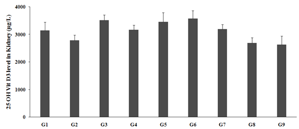
Figure 1: The effect of the test formulation on the level of 25-Hydroxy Vitamin D3 in kidney homogenate of Sprague Dawley rats. G: Group; G1: Normal control (0.5% CMC); G2: Disease control (VDD: Vitamin D3 deficient diet + 0.5% CMC); G3: Reference item (VDD + Calcitriol); G4: (VDD + untreated test formulation); G5: (VDD + Biofield Energy Treated test formulation); G6: (VDD + Biofield Energy Treatment per se to animals from day -15; G7: (VDD + Biofield Energy Treated test formulation from day -15); G8: (VDD + Biofield Energy Treatment per se plus Biofield Energy Treated test formulation from day -15), and G9: (VDD + Biofield Energy Treatment per se animals plus untreated test formulation). Values are presented as mean ± SEM (n=6).
The Effect of the Test Formulation on Kidney 1,25 Dihydroxy Vitamin D3
1, 25-dihydroxyvitamin D3 is the metabolite of vitamin D, which is one of the most biologically active form, responsible in bone resorption and intestinal absorption of calcium and phosphorus [46]. The renal formation of 1, 25-(OH)2D3 from its endogenous precursor, 25-hydroxyvitamin D3 (25-OH Vit. D3) is catalyzed by 25-Hydroxy vitamin-D3-la-hydroxylase (la- hydroxylase), an enzyme whose activity can be suppressed by 1,25-(OH)2D3 and stimulated by parathyroid hormone (PTH) [47]. The experimental data and comparison results of the test formulation on kidney 1, 25 Hydroxy vitamin D3 are presented in Figure 2. 1, 25 (OH)2 vitamin D3 level in the kidney with vitamin D3 deficient diet (G2) was 9.59 ± 0.71 ng/mL, which was significantly decreased by 18.5% as compared to the normal control (G1, 11.77 ± 0.92 ng/mL). Calcitriol (G3) group showed significantly increased the kidney 1,25 (OH)2 vitamin D3 level (10.41 ± 0.31 ng/mL) by 8.5% as compared to the G2. G4 group showed significantly decreased the kidney 1,25 (OH)2 vitamin D3 level (9.72 ± 0.72 ng/mL) by 1.2% as compared to the G2. G5 and G6 groups showed significant increased kidney 1,25 (OH)2 vitamin D3 level by 3.8% and 55%, respectively as compared with the G2. However, G7, G8, and G9 groups showed decreased kidney 1,25 (OH)2 vitamin D3 level by 5.1% and 54.2%, respectively as compared with the G2 group. The overall data suggested that preventive measures taken in the experimental groups (-15 days) showed better response as compared with the normal groups. Thus, it can be assumed that Biofield Energy Treatment per se would be the best alternative preventive approach towards any bone disorders (Figure 2).
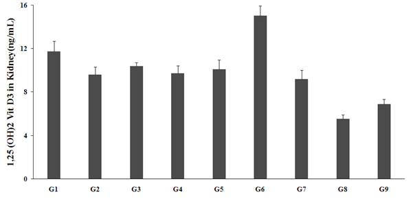
Figure 2: The effect of the test formulation on the level of 1, 25-Hydroxy Vitamin D3 in kidney homogenate of Sprague Dawley rats. G: Group; G1: Normal control (0.5% CMC); G2: Disease control (VDD: Vitamin D3 deficient diet + 0.5% CMC); G3: Reference item (VDD + Calcitriol); G4: (VDD + untreated test formulation); G5: (VDD + Biofield Energy Treated test formulation); G6: (VDD + Biofield Energy Treatment per se to animals from day -15; G7: (VDD + Biofield Energy Treated test formulation from day -15); G8: (VDD + Biofield Energy Treatment per se plus Biofield Energy Treated test formulation from day -15), and G9: (VDD + Biofield Energy Treatment per se animals plus untreated test formulation). Values are presented as mean ± SEM (n=6).
The Effect of the Test Formulation on kidney mRNA Expression for CYP24A
presented in Figure 3. CYP24A expression in the vitamin D3 deficient diet (G2) was 31.14 ± 0.51 AU decreased by 0.5% as compared to the normal control (G1). Calcitriol treatment showed significantly decreased kidney CYP24A expression (27.71 ± 0.29AU) by 11% as compared to the G2. G4 group showed significantly decreased the kidney CYP24A expression (27.71 ± 0.36AU) by 11% as compared to the G2. G5 group showed reduced level by 3.1%, while increased expression of kidney CYP24A expression by 13.9% as compared with the G4. Overall, the data suggested that the Biofield Energy Treatment per se significantly improved the enzyme expression in kidney in all the experimental preventive treatment groups as compared with the normal treatment approach (Figure 3).
CYP24A is one of the important enzyme that play a vital role in calcium homeostasis and the vitamin D endocrine system by regulating the level of vitamin D3, as it catalyzes the conversion of both 25-OH-D3 and 1,25-(OH)2D3 into 24-hydroxylated products, which are targeted for excretion using specialized pathways. CYP24A1 has been clearly established as the key enzyme responsible for vitamin D catabolism [48]. Severe infantile hypercalcemia was reported due to the abnormal functioning of CYP24A1 [49]. The experimental data and comparison results of the test formulation on kidney mRNA expression for CYP24A are
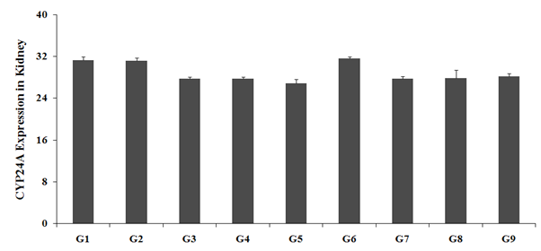
Figure 3: The effect of the test formulation on the level of kidney mRNA Expression for CYP24A in kidney homogenate of Sprague Dawley rats. G: Group; G1: Normal control (0.5% CMC); G2: Disease control (VDD: Vitamin D3 deficient diet + 0.5% CMC); G3: Reference item (VDD + Calcitriol); G4: (VDD + untreated test formulation); G5: (VDD + Biofield Energy Treated test formulation); G6: (VDD + Biofield Energy Treatment per se to animals from day -15; G7: (VDD + Biofield Energy Treated test formulation from day -15); G8: (VDD + Biofield Energy Treatment per se plus Biofield Energy Treated test formulation from day -15), and G9: (VDD + Biofield Energy Treatment per se animals plus untreated test formulation). Values are presented as mean ± SEM (n=6).
The Effect of the Test Formulation on kidney mRNA Expression for CYP27B
A CYP27B expression in the kidney has found an important role in calcitonin pathway in the kidney and in extra-renal tissues [50, 51]. Thus, the experimental data was reported and compared in animal model and is presented in Figure 4. The results suggested that vitamin D3 deficient diet (G2) was found to be 33.29 ± 0.29 AU, which was decreased by 8.3% as compared to the normal control (G1, 36.29 ± 0.36 AU). However, calcitriol treatment (G3) showed significantly decreased kidney CYP27B expression (28.86 ± 0.55 AU) by 13.3% as compared to the G2. G4 group showed significant decreased the kidney CYP27B expression (28.43 ± 1.51 AU) by 14.6% as compared to the G2. G5 showed increased the kidney CYP27B Expression by 17.6% as compared to G4. G7, G8, and G9 groups showed significant decreased expression of kidney CYP27B by 6%, 1.3%, and 13.7%, respectively as compared with the G2, while G6, G7, and G8 showed increased expression of CYP27B by 18.6%, 10.1%, and 15.6%, respectively, as compared with the G4 (Figure 4).
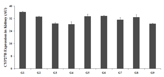
Figure 4: The effect of the test formulation on the level of kidney mRNA Expression for CYP27B in kidney homogenate of Sprague Dawley rats. G: Group; G1: Normal control (0.5% CMC); G2: Disease control (VDD: Vitamin D3 deficient diet + 0.5% CMC); G3: Reference item (VDD + Calcitriol); G4: (VDD + untreated test formulation); G5: (VDD + Biofield Energy Treated test formulation); G6: (VDD + Biofield Energy Treatment per se to animals from day -15; G7: (VDD + Biofield Energy Treated test formulation from day -15); G8: (VDD + Biofield Energy Treatment per se plus Biofield Energy Treated test formulation from day -15), and G9: (VDD + Biofield Energy Treatment per se animals plus untreated test formulation). Values are presented as mean ± SEM (n=6).
The Effect of the Test Formulation on kidney VDR expression The importance of vitamin D nuclear receptor (VDR) regulates gene expressions that are needed for calcium and phosphate homeostasis, cellular proliferation and differentiation, and immune response, largely in a ligand- dependent manner [53, 54]. VDR mediate the biological role of vitamin D and its expression was reported in most of the tissues such as bone, kidney, thyroid, and intestine, liver, kidney, and adipose tissue along with major organs [55]. VDR level in the kidney of rats was studies and is presented in Figure 5. Vitamin D3 deficient diet (G2) group showed level of VDR 15.66 ± 2.37 ng/mL, which was significantly decreased by 64.4% as compared to the normal control (G1, 44 ± 9.27 ng/mL). Calcitriol treatment (G3), treatment showed an increased the kidney VDR level (16.50 ± 2.42 ng/mL) by 5.4% as compared to the G2. G4, G6, and G7 groups showed an increased the kidney VDR level by 16%, 69.5%, 31%, and 8%, respectively as compared with the G2. However, kidney VDR level was significantly increased by 46.1% and 12.9% in G6 and G7 groups, respectively as compared with the G4 group. Overall, the data suggested that the Biofield Energy Treatment per se significantly improved the VDR expression in kidney in all the experimental groups along with the preventive treatment groups (Figure 5).
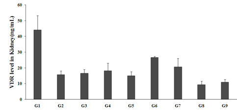
Figure 5: The effect of the test formulation on the level of kidney VDR level in kidney homogenate of Sprague Dawley rats. G: Group; G1: Normal control (0.5% CMC); G2: Disease control (VDD: Vitamin D3 deficient diet + 0.5% CMC); G3: Reference item (VDD + Calcitriol); G4: (VDD + untreated test formulation); G5: (VDD + Biofield Energy Treated test formulation); G6: (VDD + Biofield Energy Treatment per se to animals from day -15; G7: (VDD + Biofield Energy Treated test formulation from day -15); G8: (VDD + Biofield Energy Treatment per se plus Biofield Energy Treated test formulation from day -15), and G9: (VDD + Biofield Energy Treatment per se animals plus untreated test formulation). Values are presented as mean ± SEM (n=6).
Thus, the present research plan defined four groups, which were considered as preventive maintenance groups viz. G6, G7, G8, and G9, where the Biofield Energy Treatment per se and/or Biofield Energy Treated Test formulation in combination was used as preventive maintenance group. The results showed the significant slowdown of the disease progression, disease related all other complications and also reduced the chances of disease susceptibility in these groups. Specifically, group G6 (preventive Biofield Energy Treatment group per se) showed the best results as a prophylactic/preventive treatment group compared to the other groups. Based on the overall data, it suggests that the Biofield Energy Healing Therapy was found to be most effective and benefited in order to prevent and protect from the occurrence of any type of bone related diseases in rat model. It indicated that Biofield Energy Treatment can act as a preventive maintenance therapy to slow down the disease progression and disease related complications of the existing aliments that will ultimately improve the overall health and quality of life in human.
Conclusions
The present animal experimental study revealed the significance of Biofield Energy Treated test formulation and Biofield Energy per se on the vitamin D metabolites in kidney tissues. The Biofield Energy Treated test formulation (G5), Biofield Energy Treatment per se (G6), and 15-days pre- treatment of the Biofield Energy Treated test formulation (G7) groups were reported with an increased kidney 25-OH vitamin D3 level by 21%, 22.9%, and 11.5%, respectively as compared with the disease control group (G2). Further, G6 group was reported with an increased level of renal formation of 1,25-(OH)2D3 by 55% as compared with the G4. CYP24A expression in kidney was increased by 13.9% in the G6 group, while CYP27B expression in kidney was significantly increased by 17.6%, 18.6%, 10.1%, and 15.6% in the G5, G6, G7, and G8 groups, respectively as compared with the G4 group. VDR expression was significantly improved by 69.5% and 31% in the G6 and G7 groups, respectively as compared with the G2 group. Overall, the results showed significant improvement of the availability of vitamin D3 metabolites, that could be helpful to slow down the disease progression and disease related complications of the overall animal’s health. It can be concluded Biofield Energy Healing Treatment (the Trivedi Effect®) per se showed best results with respect to different biomarker parameters in the preventive treatment approach (-15 days) as compared to the other preventive maintenance groups (G7, G8, and G9) in rat model study. These data suggested that Biofield Energy Treatment per se and/or Biofield Energy Treated Test formulation in combination would be the best treatment strategies in order to prevent and protect from the occurrence of any type of diseases. Therefore, The Biofield Energy Treatment might act as a preventive maintenance therapy in order to cure, or full restoration of health or improve the overall health and quality of life in human. This therapy might also reduce the severity of any type of acute/ chronic disease (auto-immune related and inflammatory disorders) progression rate and can be given in both before and after the manifestation of any disease symptoms in healthy, unhealthy, and ill peoples such as many thyroid disorders such hyperthyroidism, Goiter, Thyroid nodules, Thyroid cancer, Thyroid hormone resistance, Hashimoto’s thyroiditis, Anaplastic Thyroid Cancer, Hypothyroidism, De Quervain’s Thyroiditis, Medullary Thyroid Cancer, Follicular Thyroid Cancer, Papillary Thyroid Cancer, Silent Thyroiditis, Graves’ Disease, Thyroid Cancer, Hurthle Cell Thyroid Cancer, and Thyroiditis. Overall, the data suggested the Biofield Energy Treated test formulation and Biofield Energy Treatment per se in showed significant action on thyroid gland with respect to biomarkers, as a Complementary and Alternative Medicine (CAM). This test formulation also can be used against Lupus, Fibromyalgia, Addison Disease, Multiple Sclerosis, Myasthenia Gravis, Aplastic Anemia, Psoriasis, Rheumatoid Arthritis, Crohn’s Disease, Vitiligo, Chronic Fatigue Syndrome and Alopecia Areata, as well as various inflammatory disorders such as Ulcerative Colitis, Dermatitis, Hepatitis, Diverticulitis, Mental Disorders, Parkinson’s and Other Movement Disorders, Stroke and Transient Ischemic Attack (TIA), and in the improvement of overall health and quality of life.
Acknowledgements
The authors are grateful to Dabur Research Foundation,
Trivedi Science, Trivedi Global, Inc., and Trivedi Master Wellness for the assistance and support during the work.
References
-
Nair R, Maseeh A (2012) Vitamin D: The “sunshine” vitamin. J Pharmacol Pharmacother 3(2): 118- 126.
-
Holick MF (2007) Vitamin D deficiency. N Engl J Med 357: 266-281.
-
Gordon CM, DePeter KC, Feldman HA, Grace E, Emans SJ (2004) Prevalence of vitamin D deficiency among healthy adolescents. Arch Pediatr Adolesc Med 158(6): 531-537.
-
Melamed ML, Michos ED, Post W, Astor B (2008) 25-hydroxyvitamin D levels and the risk of mortality in the general population. Arch Intern Med 168(15): 1629- 1637.
-
Bikle DD (2014) Vitamin D metabolism, mechanism of action, and clinical applications. Chem Biol 21(3): 319- 329.
-
Christakos S, Ajibade DV, Dhawan P, Fechner AJ, Mady LJ (2010) Vitamin D: metabolism. Endocrinol Metab Clin North Am 39(2): 243-253.
-
Hollis BW, Wagner CL, Drezner MK, Binkley NC (2007) Circulating vitamin D3 and 25-hydroxyvitamin D in humans: An important tool to define adequate nutritional vitamin D status. J Steroid Biochem Mol Biol 103(3-5): 631-634.
-
Zhu JG, Ochalek JT, Kaufmann M, Jones G, Deluca HF (2013) CYP2R1 is a major, but not exclusive, contributor to 25-hydroxyvitamin D production in vivo. Proc Natl Acad Sci U S A 110(39): 15650-15655.
-
Bikle DD (2009) Extra renal synthesis of 1, 25 dihydroxyvitamin D and its Health Implications. Clin Rev in Bone and Min Metab 7: 114-125.
-
Franca Gois PH, Wolley M, Ranganathan D, Seguro AC (2018) Vitamin D Deficiency in Chronic Kidney Disease: Recent Evidence and Controversies. Int J Environ Res Public Health 15(8):1773.
-
Williams S, Malatesta K, Norris K (2009) Vitamin D and chronic kidney disease. Ethn Dis 19(4 Suppl 5): S5- S11.
-
Jean G, Souberbielle JC, Chazot C (2017) Vitamin D in Chronic Kidney Disease and Dialysis Patients. Nutrients 9(4): 328.
-
Restrepo Valencia CA, Aguirre Arango JV (2019) Vitamin D (25(OH)D) in patients with chronic kidney disease stages 2-5. Colomb Med (Cali) 50(1): 49.
-
Al-Badr W, Martin KJ (2008) Vitamin D and kidney disease. CJASN 3(5): 1555-1560.
-
Dusso AS, Brown AJ, Slatopolsky E (2005) Vitamin D. Am J Physiol Renal Physiol 289: 8-28.
-
Hilpert J, Wogensen L, Thykjaer T, Wellner M, Schlichting U, et al. (2002) Expression profiling confirms the role of endocytic receptor megalin in renal vitamin D3 metabolism. Kidney Int 62(5): 1672-1681.
-
Willnow TE, Nykjaer A (2002) Pathways for kidney- specific uptake of the steroid hormone 25-hydroxyvitamin D3. Curr Opin Lipidol 13(3): 255-260.
-
Byrne JH, Voogt M, Turner KM, Eyles DW, McGrath JJ, et al. (2013) The impact of adult vitamin D deficiency on behaviour and brain function in male Sprague-Dawley rats. PLoS One 8(8): e71593.
-
Rayman MP (2000) The importance of selenium to human health. Lancet 356(9225): 233-241.
-
Beard JL, Connor JR (2003) Iron status and neural functioning. Ann Rev Nutr 23: 41-58.
-
Peres FF, Lima AC, Hallak JEC, Crippa JA, Silva RH, et al. (2018) Cannabidiol as a Promising Strategy to Treat and Prevent Movement Disorders? Front Pharmacol 9: 482.
-
Nagarkatti P, Pandey R, Rieder SA, Hegde VL, Nagarkatti M (2009) Cannabinoids as novel anti-inflammatory drugs. Future Med Chem 1(7): 1333-1349.
-
Kang S, Min H (2012) Ginseng, the ‘Immunity Boost’: the effects of Panax ginseng on immune system. J Ginseng Res 36(4): 354-368.
-
Maizes V, Rakel D, Niemiec C (2009) Integrative medicine and patient-centered care. Explore (NY) 5(5): 277-289.
-
Bischof M, Del Giudice E (2013) Communication and the emergence of collective behavior in living organisms: A quantum approach. Mol Biol Int 2013: 987549.
-
Cassidy CM (2004) What does it mean to practice an energy medicine? J Altern Complement Med 10(1): 79- 81.
-
Barnes PM, Bloom B, Nahin RL (2008) Complementary and alternative medicine use among adults and children: United States, 2007. Natl Health Stat Report 12: 1-23.
-
Fan K wai (2005) National Center for Complementary and Alternative Medicine Website. J Med Libr Assoc 93: 410-412.
-
Wisneski LA, Anderson L (2009) The Scientific Basis of Integrative Medicine. Boca Raton, CRC Press, pp: 205.
-
Trivedi MK, Tallapragada RM (2008) A transcendental to changing metal powder characteristics. Met Powder Rep 63: 22-29, 31.
-
Trivedi MK, Nayak G, Patil S, Tallapragada RM, Latiyal O (2015) Studies of the atomic and crystalline characteristics of ceramic oxide nano powders after biofield treatment. IndEng Manage 4(3): 161.
-
Trivedi MK, Branton A, Trivedi D, Nayak G, Jana S, et al. (2015) Morphological characterization, quality, yield and DNA fingerprinting of biofield energy treated alphonso mango (Mangifera indica L.). Journal of Food and Nutrition Sciences 3(6): 245-250.
-
Trivedi MK, Branton A, Trivedi D, Nayak G, Charan S, Jana S (2015) Phenotyping and 16S rDNA analysis after biofield treatment on Citrobacter braakii: A urinary pathogen. J Clin Med Genom 3(1): 129.
-
Trivedi MK, Patil S, Shettigar H, Mondal SC, Jana S (2015) Evaluation of biofield modality on viral load of Hepatitis B and C viruses. J Antivir Antiretrovir 7(3): 083-088.
-
Nayak G, Altekar N (2015) Effect of biofield treatment on plant growth and adaptation. J Environ Health Sci 1(2): 1-9.
-
Branton A, Jana S (2017) The influence of energy of consciousness healing treatment on low bioavailable resveratrol in male Sprague Dawley rats. International Journal of Clinical and Developmental Anatomy 3(3): 9-15.
-
Branton A, Jana S (2017) The use of novel and unique biofield energy healing treatment for the improvement of poorly bioavailable compound, berberine in male Sprague Dawley rats. American Journal of Clinical and Experimental Medicine 5(4): 138-144.
-
Kinney JP, Trivedi MK, Branton A, Trivedi D, Nayak G, et al. (2017) Overall skin health potential of the biofield energy healing based herbomineral formulation using various skin parameters. American Journal of Life Sciences 5(2): 65-74.
-
Singh J, Trivedi MK, Branton A, Trivedi D, Nayak G, et al. (2017) Consciousness energy healing treatment based herbomineral formulation: A safe and effective approach for skin health. American Journal of Pharmacology and Phytotherapy 2(1): 1-10.
-
Trivedi MK, Branton A, Trivedi D, Nayak G, Plikerd WD, et al. (2017) A Systematic study of the biofield energy healing treatment on physicochemical, thermal, structural, and behavioral properties of magnesium gluconate. International Journal of Bioorganic Chemistry 2(3): 135-145.
-
Trivedi MK, Patil S, Shettigar H, Mondal SC, Jana S (2015) The potential impact of biofield treatment on human brain tumor cells: A time-lapse video microscopy. J Integr Oncol 4(3): 141.
-
Anagnos D, Trivedi K, Branton A, Trivedi D, Nayak G, Mondal SC, Jana S (2018) Influence of biofield treated vitamin D3 on proliferation, differentiation, and maturation of bone-related parameters in MG-63 cell- line. International Journal of Biomedical Engineering and Clinical Science 4(1): 6-14.
-
Lee AC, Trivedi K, Branton A, Trivedi D, Nayak G, et al. (2018) The potential benefits of biofield energy treated vitamin D3 on bone mineralization in human bone osteosarcoma cells (MG-63). International Journal of Nutrition and Food Sciences 7(1): 30-38.
-
Khazai N, Judd SE, Tangpricha V (2008) Calcium and vitamin D: skeletal and extraskeletal health. Curr Rheumatol Rep 10(2): 110-117.
-
DeLuca HF (1980) The control of calcium and phosphorus metabolism by the vitamin D endocrine system. Ann N Y Acad Sci 355: 1-17.
-
Christakos S, Ajibade DV, Dhawan P, Fechner AJ, Mady LJ (2010) Vitamin D: metabolism. Endocrinol Metab Clin North Am 39(2): 243-253.
-
Dusso AS (2011) Kidney disease and vitamin D levels: 25-hydroxyvitamin D, 1,25-dihydroxyvitamin D, and VDR activation. Kidney Int Suppl 1(4): 136-141.
-
Anderson PH, May BK, Morris HA (2003) Vitamin D metabolism: new concepts and clinical implications. Clin Biochem Rev 24(1): 13-26.
-
Dauber A, Nguyen TT, Sochett E, David ECC, Horst R, et al. (2012) Genetic defect in CYP24A1, the vitamin D 24-hydroxylase gene, in a patient with severe infantile hypercalcemia. J Clin Endocrinol Metab 97(2): E268– E274.
-
Anderson PH, O’Loughlin PD, May BK, Morris HA (2005) Modulation of CYP27B1 and CYP24 mRNA expression in bone is independent of circulating 1,25(OH)2D3 levels. Bone 36(4): 654-662.
-
Chanakul A, Zhang MY, Louw A, Armbrecht HJ, Miller WL, et al. (2013) FGF-23 regulates CYP27B1 transcription in the kidney and in extra-renal tissues. PLoS One 8(9): e72816.
-
Takeyama K, Kitanaka S, Sato T, Kobori M, Yanagisawa J, et al. (1997) “25-Hydroxyvitamin D3 1alpha-hydroxylase and vitamin D synthesis”. Science 277 (5333): 1827- 1830.
-
Malloy PJ, Feldman D (2011) The role of vitamin D receptor mutations in the development of alopecia. Mol Cell Endocrinol 347(1-2): 90-96.
-
Ryan JW, Anderson PH, Turner AG, Morris HA (2013) Vitamin D activities and metabolic bone disease. Clin Chim Acta 425: 148-152.
-
Wang Y, Borchert ML, DeLuca HF (2012) Identification of the vitamin D receptor in various cells of the mouse kidney. Kidney Int 81(10): 993-1001.
- Effects of 5-HTP and Melatonin on the Sleep Cycle of Medical Students
- Adsorption of Bisphenol A on NH4OH- Modified Rice Husk and Sugar Cane Bagasse Biochar
- Comparative Assessment of the Reinforcement Efficiency of Palm Fruit Fibre and Coconut Fibre in High Density Polyethylene (HDPE) Matrix Composite
- Importance of Bio Compounds Naturally Present in Food with Functionality in Animal Metabolism
- Sub-Acute Study on the Cardiotoxic Effects of Monosodium Glutamate Ingestion in Albino Rat
- Weight Management and Its Natural Solutions: A Review