Black Tea (Camellia sinensis) Extract Induced Changes on Placenta can Alter Fetal and Neonatal Bone Health in Experimental Animal Model
Tea (Camellia sinensis) being the most consumed beverage worldwide. The safety evaluation of tea needs to be monitored during pregnancy, prenatal and postnatal developmental period beside its beneficial roles toward health and disease. Retardation of growth of fetus and neonates are common in preeclampsia. Present study was to evaluate the role of Black Tea extract (BTE) on placental and apoptotic markers in pregnant Wister albino rats and to correlate it the growth of fetus and pups. Among three experimental groups, Group 1 was pregnant female rats treated with saline, were the control group. Group 2 and Group 3 were pregnant female rats treated with 50 mg and 100 mg BTE/kg/day, p.o. respectively throughout prenatal and postnatal periods. Expressions of BMP-7, MMP-2 and VEGFR2 in placenta were examined by flow cytometry; Bax, Bcl-2 and caspase-3 expressions in uteri and placenta were observed by IHC. Bone health of fetus and pup were checked by histology, bone-cartilage double staining and estimation of bone mineral density by ICP-MS. Experimental data were subjected to the ANOVA; expressed as mean ± standard deviation with significance (P < 0.05) between the controls and the treated groups (n = 6). BTE increased the level of MMP-2, Bax and caspase-3; decreased the level of BMP-7 in placenta. In fetus and pups, BTE significantly decreased the concentration of Ca2+, P, Mg2+ and Zn2+ in bone and decreased the rate of ossification were observed. This study confirmed BTE induced preeclampsia retarded the fetal and neonatal bone health in experimental animal model.
Introduction
Tea (Camellia sinensis) is native to the different Asian countries like China, India, Laos, Thailand, Vietnam, and Myanmar [1]. Tea is one of the most widely consumed beverages in the world, with a global market comprising of four major zones: Asia-Pacific, Europe, North America and Africa [2, 3]. The tea is second only to water in terms of worldwide consumption and presently tea is cultivated in over thirty countries around the world [4, 5]. Tea is classified into three types; green tea, black tea and oolong tea. The classification of tea is based on the fermentation and oxidation of the polyphenols in the tea leaves during production [6]. Green
tea is the non-fermented form of tea, in which the oxidation of the tea polyphenols called catechins is prevented and thus, most of the catechins are preserved during its processing. Black tea and oolong tea are respectively fully fermented and semi-fermented tea leaves. In black tea and oolong tea, leaf polyphenolics are allowed for aerobic oxidation and the catechins are enzymatically catalysed to form theaflavins and thearubigins [4, 7]. In case of black tea the reaction is carried out to maximize the oxidation (fermentation) but for oolong tea reaction is stopped usually half-way before it is completely oxidised. There are two main types of black tea; orthodox (rolling) black tea and CTC (crushing, tearing and curling) black tea, produced through various stages including withering, rolling, drying and grading [8]. Tea and its bioactive components have the potential in disease prevention and are effective in therapy. Both green tea and black tea are cardio protective [9, 10], antioxidant and anti- inflammatory [11, 12], has anti-cancer effects [13, 14], anti- obese effects [15], neuroprotective effects [16]. Despite the increasing demand for tea and its active constituents, few studies have reported their safety. Tea has numerous beneficial roles towards health and disease but its safety evaluation during pregnancy and prenatal as well as in postnatal developmental period need to be monitored. Very few studies have been reported regarding tea extract consumption and its effect during pregnancy in animal models [17, 18]. Pu-erh black tea a highly fermented version of black tea is associated with development of fetal toxicity at a high concentration [19]. Preeclampsia is a disease of late pregnancy characterized by increased maternal blood pressure, proteinuria, increase in pro-inflammatory cytokine and decrease in anti-inflammatory cytokine [20, 21]. According to Dey, et al. [22] black tea induced preeclampsia in experimental Wister albino rat [22]. In the present study, an attempt has been made to assess the effect of varying doses of black tea extract (BTE) on some placental and apoptotic markers that caused preeclamsia in pregnant rats which may further leads to retardation of growth in fetus and pups.
Materials and Methods
Chemicals
Absolute alcohol (ethanol) and Methanol (Merck, India), Alcian blue 8GX (Sigma-Aldrich, USA), Alizarin Red S (Sigma-Aldrich, USA), Anti-rat Alexa Fluor 488-MMP- 2 (Novus Biologicals, USA) for FACS, Anti-rat Bax, Bcl2, caspase-3 (Santa Cruz Biotechnology, United States) for immunohistochemistry, Anti-rat FITC BMP-7 (Milli-Mark, USA) for FACS, Anti-rat PE VEGFR2 (BioLegend, USA) for FACS, Avitin-Biotin Conjugate (Thermo Fisher Scientific, United States) for immunohistochemistry, Benzyl alcohol (Milli-Mark, India), Biotin-conjugated anti-rat secondary antibodies (Thermo Fisher Scientific, United States) for immunohistochemistry, Collagenase (type IV) (Himedia, India), DAB substrate and diluent (Thermo Fisher Scientific, United States) for immunohistochemistry, DPX (LOBA Chemie, India), Di-sodium hydrogen phosphate (SRL, India), Sodium di-hydrogen phosphate (SRL, India), Eosin (Sigma, USA), Fetal calf serum (Sigma-Aldrich, USA), Formaldehyde solution 37-41% w/v (Merck, India), Glacial Acetic Acid (Merck, India), Glycerol anhydrous (Milli-Mark, India), Hematoxylin (Merck, Germany), Hydrogen peroxide (Milli- Mark, India), Paraffin wax 56-58°C (Merck, India), Nitric acid (Milli-Mark, India), Potassium hydroxide (Merck, India), Salt mixture H.M.W. (SRL, India), Sodium chloride (SRL, India), Tri-sodium citrate (Merck, India), Trizma® base (SRL, India), Tween-20 (Milli-Mark, India), Xylene (Merck, India).
Animals
Male (150±10 g) and female (120±10 g) wister albino rats were obtained from the enlisted supplier of CPCSEA (Committee for the Purpose of Control and Supervision of Experiments on Animal), India. Rats were kept in polypropylene cages (421×290×190mm) at controlled temperature (25±2°), light condition (12h light and dark cycle) and relative humidity (65±5%). The animals were provided with pellet diet, green vegetables, gram and water ad libitum. All animals for this experiment were kept in CPCSEA approved animal house (vide F. No. - 25/250/2012- AWD, dated 26.2.2014) of Maulana Azad College, Kolkata. Experiments described in this study were done by following the guideline of the CPCSEA, Government of India.
Collection of Black Tea
Fresh black tea (C.T.C., Assam) was purchased from authenticated tea supplier M/S. Subodh Brothers Pvt. Ltd., Kolkata-700012, India.
• Preparation of black tea extract and treatment schedule Black tea extract (BTE) was prepared after Dey, et al. [22]. First, 1 g black tea was added into 100 ml of boiled drinking water, was kept covered for 5 minutes, filtered by a tea strainer. The dry weight of 100 ml freshly prepared BTE was calculated by evaporating the water from which is equivalent to one cup of black tea liquor. Dry weight of BTE/ml was also calculated in the same way by adding 1 g of black tea in 6 ml boiled drinking water. 50 mg and 100 mg BTE/kg b.w./ day, p.o. were the two doses selected for treatment which are equivalent to 5 cup and 10 cup BTE respectively in human considering 60 kg as average body weight of an adult human. 1 g of black tea was added in 6 ml boiled drinking water, was kept covered for 5 minutes, filtered and cooled down to 40°C.
200 µl and 400 µl BTE from this liquor was administered orally to achieve respective dose of 50 mg and 100 mg BTE/ kg b.w./day. The doses were administered orally by the help of oral gavage to the pregnant rats throughout their prenatal (21 days) and postnatal periods of time (21days).
Experimental Design
The pregnant rats were selected by pairing a pro-estrous female overnight with two male rats of proven fertility and determining the vaginal sperm count on the following day (9:00 to 9:30 a.m.) using an improved Neubauer haemocytometer. Rats with vaginal sperm counts >45×106/ ml were selected and considered as at day 0 of pregnancy. For this study three animal groups were chosen (n=8 per group). Group 1 was the control group where the pregnant female rats were treated with saline. Group 2 and Group 3 were pregnant female rats treated with 50 mg and 100 mg BTE/kg body weight/day, p.o. respectively. Both the doses of BTE were administered in the pregnant rats throughout their prenatal (21 days) and postnatal periods (21days) of time. All three groups of rats were provided with pellet diet, gram, green vegetables and drinking water ad libitum.
Isolation of Placenta and Uterus
Pregnant rats (on day 20) of three different groups were euthanized according to CPCSEA approved procedures. Uterine horns were picked up, collected by using scissors at each distal end, placed in a petri dish filled with PBS and place on ice, washed several times with PBS before isolating the placenta. The endometrial tissue was carefully peeled out surrounding the embryo conceptuses. Isolated conceptuses were placed in a new petri dish with PBS on ice and continue until all conceptuses have been freed from the endometrium. Conceptuses were transferred to a new petri dish with PBS placed under the microscope and with two forceps peel the decidua away from the placenta. Yolk sacs, excess giant cell tissue at the edges of the placenta were removed in order to minimize cell clumping during preparation of single-cell suspension. Placentas for FACS analysis were collected in a 15-ml Falcon tube, with PBS+5% FCS and on ice [23]. Uteri and placenta of different groups were fixed in 10% neutral buffered formalin for immunohistochemistry.
• Study of placental BMP-7, MMP-2 and VEGFR2 positive cells population by flow cytometry
0.1% Collagenase solution in PBS with 10% FCS was prepared and was added into placenta (5ml/placenta). 16-G needle fitted on a 5-ml syringe was used to mechanically disrupt the tissue by passaging the collagenase solution and placenta through the needle 3 times. It was repeated with an 18-G needle. The mixture was placed in a 37°C, 5% CO2 incubator for 45 minutes. After that the cell solution was passed through a 20-G needle, and incubate for an additional 45 minutes, in 37°C. After the 1.5 hr total incubation in collagenase, cell solution was passed through 22-G and 25-G needles 3 times with each needle [23]. Placental digests were filtered through a 40µm nylon cell-strainer and red blood cells were removed by centrifuging the placental digests at 4°C for 5 minutes at 2000 rpm using RBC lysis buffer solution. Next cells were washed, counted and re-suspended in PBS to make single-cell suspension. Accurate cell count was taken and then cells were divided into aliquots having 106cells/100 µl of cell staining buffer (CSB), containing 3% foetal calf serum (FCS). Further the cells were incubated with the titrated amount of anti-rat PE (Phycoerythrin dye)-CD309 (VEGFR2, Flk-1), FITC (Fluorescein isothiocyanate dye) BMP-7, Alexa Fluor 488 – MMP-2, fluorochrome-conjugated primary antibody in the dark, and incubated for 1 h [24]. Finally the cells were washed and the pellets were resuspended in 100µl CSB and were analyzed using BD FACSVerse flow cytometer and BD FACSuite TM Software.
• Immunohistochemistry of placental Bax, Bcl-2 and caspase-3 For immunohistochemistry (IHC) Uteri and placenta of different groups were fixed in 10% neutral buffered formalin. Tissues were then dehydrated in graded (50- 100%) ethanol followed by clearing in xylene. Paraffin (56°C-58°C) embedding was done at 58±1°C for 4 h, followed by paraffin block preparation. 4μm thick paraffin embedded section was cut with a rotary microtome (Weswox model MT-1090, India). Tissue sections were mounted on poly- L-Lysine coated slides. Sections were deparaffinised, dehydrated through graded alcohols, antigen retrival was done by 10mM sodium citrate and endogenous peroxidase was quenched by 3% hydrogen peroxide (H2O2). After blocking with 1% foetal calf serum (FCS) in tris buffer saline (TBS), the sections were incubated in a humid chamber overnight at 4°C with primary antibodies like anti-rat Bax, Bcl2, caspase-3 (Santa Cruz Biotechnology, United States). After washing in wash buffer (1% Tween 20 in TBS or 1X TBST), sections were incubated in biotin-conjugated anti- rat secondary antibodies diluted in Tris-buffered saline (TBS) for 2 hours at room temperature. After washing in 1X TBST sections were incubated in Avidin-Biotin Conjugate (ABC) for 30 minutes. Immunoreactivity was detected using a DAB system. Sections were then counterstained briefly in hematoxylin, dehydrated through graded alcohols, cleared in xylene, and cover-slipped with DPX [25, 26]. Images were captured and changes were observed with bright field microscope (ZEISS, Germany) and photographs were taken by using ZEISS AxioCam ICc1 and Zen software (Zen2 lite) at 100X magnification. Quantification of Bax, Bcl2, caspase 3
positive cells was calculated by ImageJ software. Percentage of placental cells positive for Bax, Bcl-2 and caspase 3 was calculated separately by ImageJ. An average of 20 fields was observed for three different groups. The ratios of the Bax : Bcl-2 was calculated.
Bone and Cartilage Double Staining of Pups
Day 0 pups of three different groups were collected etherized and were put into 4% NaCl solution for overnight at 4°C. On next day fetuses were skinned and eviscerated, the cervical and dorsal muscles gently removed. The specimens were immediately placed in the acid staining solution (pH 2.8) for at least 24 hr at room temperature. It was then dehydrated in ethanol 96% for at least 6 hr. Maceration of soft tissues was performed by placing specimens in the basic staining solution for 30 hr at room temperature, while renewing the solution at least three times. Clearing and hardening was performed by placing specimens in the cleaning solution for at least 8 hr. Conservation of double- stained fetuses was performed in a 1:1 ethanol 70% and glycerin mixture [27].
The acid staining solution (pH 2.8) made up of 5 parts 0.14% Alcian blue (dissolved in 70% ethanol, filtered),1 part of 0.12% Alizarin red S (dissolved in 96% ethanol, filtered), 8 parts glacial acetic acid and 50 parts 70% ethanol. On the other hand the basic staining solution consisted of 250 parts 0.7% KOH (dissolved in distilled water) and 1part 0.5% Alizarin red S (dissolved in distilled water, filtered). The clearing solution consisted of 2 parts 70% ethanol, 2 parts glycerin and 1 part benzyl alcohol.
Estimation of Bone Mineral Density of Pups
Left femur was collected from day 20 foetus and day 21 pups of three different groups. Bone marrow was cleaned out from the bones. Bone (left femur) ash was prepared in a muffle furnace (700°C×6 h) [28] and dissolved in 5% HNO3. Bone minerals (Ca2+, P, Mg2+ and Zn2+) were measured by using Inductively Coupled Plasma – Mass Spectroscopy (ICP- MS) (ThermoFisher-Scientific X-series 2, Massachusetts, USA). The samples were ran with respect to previously known concentrations of the elements to determine their respective values. Measurement of samples were carried using standard curves having regression values greater than 0.99.
Histology of Femur of Pups
Femurs were dissected out from day 21 pups of three different groups; bone marrow was washed out by flushing double distilled water through femur. Then the bones were suspended in osteomoll for decalcification [29]. After the tissue became soft the tissues were fixed in 10% neutral buffered formalin for 24 h. Tissues were then dehydrated in graded (50-100%) ethanol followed by clearing in xylene. Paraffin (56°C-58°C) embedding was done at 58±1 °C for 4 h, followed by paraffin block preparation. 4μm thick paraffin embedded section was cut with a rotary microtome (Weswox model MT-1090, India). Xylene was used to deparaffinise the paraffin sections, and then counter stained with haematoxyiln-eosin and was mounted in DPX with a cover slip. Images were captured and changes were observed with bright field microscope (ZEISS, Germany) and photographs were taken by using ZEISS AxioCam ICc1 and Zen software (Zen2 lite) at 100X magnification.
Statistical Analysis
The data generated on various parameters were subjected to statistical analysis were expressed as means and standard deviation (mean ± SD) or mean ± SEM with significance between the controls and the treated. Collected data were subjected to one-way analysis of variance (ANOVA) considering p-values of <0.05 were considered as significant. SPSS 17.0 software (IBM Corporation, United States) was used for statistical analysis.
Results
Expression of BMP7+VEGFR2+ and MMP2+VEGFR2+ Double Positive Cell Population
FACS analysis showed no significantly (p<0.05) changes were found in BMP7+VEGFR2+ cell population of three different experimental groups (Figure 1A,B). Interestingly, the percentage of BMP7+ cells is significantly (p<0.05) decreased in group 3 as compared to control group (Figure 1A,C). On the other hand, BTE (100 mg /kg body weight/day, p.o.) significantly (p<0.05) decreased MMP2+VEGFR2+ cell population compared to control group (Figure 2 A,B) whereas administration of BTE (100 mg /kg body weight/day, p.o.) significantly (p<0.05) increased MMP2+ cells percentage in group 3 compared to control group (Figure 2 A,C).
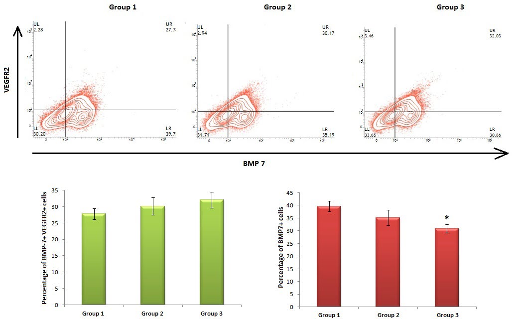
Figure 1: Expression pattern of BMP7+VEGFR2+ double positive cell population. (A) Contour plots of BMP7-VEGFR2 were obtained from the single cell suspension prepared from placenta. (B) Bar diagram representing percentage of BMP7+VEGFR2+ double positive cells. (C) Bar diagram representing percentage of BMP7+ positive cells. Data represent the mean±SD (n=6). *p< 0.05 when compared to animals of group 1. Gr. 1: control group, Gr. 2: pregnant female rats treated with BTE (50 mg BTE/ kg body weight/day, p.o.), Gr.3: pregnant female rats treated with BTE (100 mg BTE/kg body weight/day, p.o.).
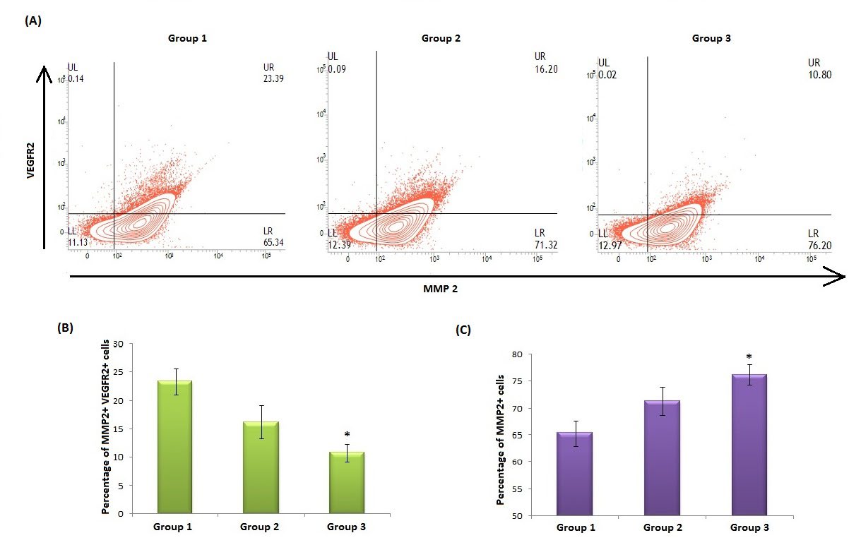
Figure 2: Expression pattern of MMP2+VEGFR2+ double positive cell population. (A) Contour plots of MMP2-VEGFR2 were obtained from the single cell suspension prepared from placenta. (B) Bar diagram representing percentage of MMP2+VEGFR2+ double positive cells. (C) Bar diagram representing percentage of MMP2+ positive cells. Data represent the mean±SD (n=6). *p< 0.05 when compared to animals of group 1. Gr. 1: control group, Gr. 2: pregnant female rats treated with BTE (50 mg BTE/ kg body weight/day, p.o.), Gr.3: pregnant female rats treated with BTE (100 mg BTE/kg body weight/day, p.o.).
Expression of Bax, Bcl-2 and Caspase-3
Immunohistochemistry showed BTE (100 mg /kg body weight/day, p.o.) increased the expression of Bax, Bcl-2 and caspase-3 in placenta and uteri compared to control group. Orange coloured dots indicate the antibody binding region. BTE (50 mg /kg body weight/day, p.o.) showed no significant changes in placenta and uteri (Figures 3 & 4).
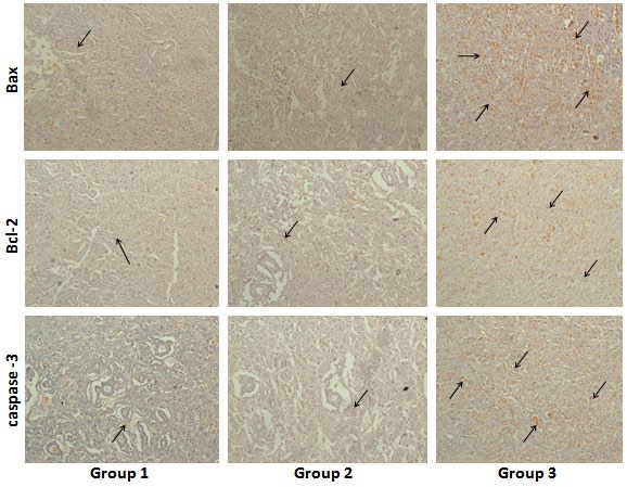
Figure 3: Localization of Bax, Bcl-2 and caspase-3 positive cells in placenta examined by immunohistochemistry (100X magnification). Gr. 1: control group, Gr. 2: pregnant female rats treated with BTE (50 mg /kg body weight/ day, p.o.), Gr.3: pregnant female rats treated with BTE (100 mg /kg body weight/day, p.o.). Black arrow indicating the expression of respective antibodies in placenta of three different groups of mothers on day 20.
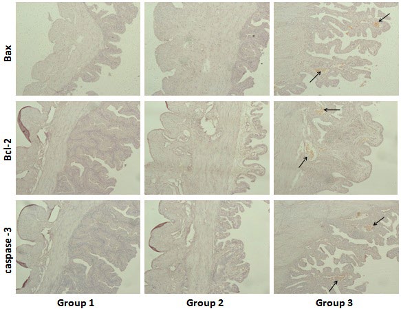
Figure 4: Localization of Bax, Bcl-2 and caspase-3 positive cells in uteri examined by immunohistochemistry (100X magnification). Gr. 1: control group, Gr. 2: pregnant female rats treated with BTE (50 mg /kg body weight/day, p.o.), Gr.3: pregnant female rats treated with BTE (100 mg / kg body weight/day, p.o.). Black arrow indicating the expression of respective antibodies in uteri of three different groups of mothers on day 20.
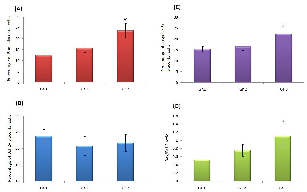
Figure 5: Quantitative expression of Bax, Bcl-2 and caspase-3 positive cells in placenta. (A) Histogram showing the percentage of Bax+ placental cell population, (B) Percentage of Bcl-2+ placental cell population, (C) Percentage of caspase-3+ placental cell population, (D) Bax/Bcl-2 ratio of placental cell population in three different groups. Data represent the mean±SD (n=6). *p< 0.05 when compared to animals of group 1. Gr. 1: control group, Gr. 2: pregnant female rats treated with BTE (50 mg /kg body weight/day, p.o.), Gr.3: pregnant female rats treated with BTE (100 mg /kg body weight/day, p.o.).
Quantitative analysis by ImageJ software showed percentage of Bax+ and caspase-3+ placental cell population were significantly (p<0.05) increased in Group 3 mother compared to Group 1 (Figure 5 A,C). There was no significant alteration in Bcl-2+ placental cell population in all experimental groups (Figure 5B). The Bax/Bcl-2 ratio was also significantly (p<0.05) increased in Group 3 placenta compared to Group 1 (Figure 5 D).
Effect of BTE on Bone and Cartilage
Bone and cartilage double staining of day 0 pups showed rate of ossification of carpus, metacurpus, phalanges of fore limbs and tarsus, metatarsus, phalanges of hind limbs was lesser in Gr.2 and Gr.3 pups compared to Gr.1 pups. An unstained gap was found between parietal bones of skull in pups of Gr.2 and Gr.3 mothers. Parietal bones were complete and no gap was found in Gr.1 pups (Figure 6).
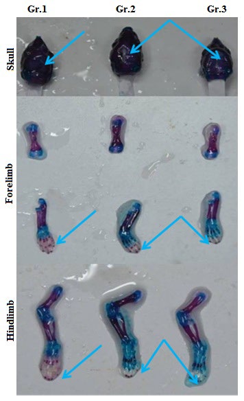
Figure 6: Bone and cartilage double staining of day 0 pups (Alcian blue- Alizarin red counterstaining). Blue colour indicates cartilaginous part and red colour indicates ossified part. Gr. 1: pups of control group, Gr. 2: pups of pregnant female rats treated with BTE (50 mg /kg body weight/day, p.o.), Gr.3: pups of pregnant female rats treated with BTE (100 mg /kg body weight/day, p.o.). Blue arrows are indicating the changes in rate of ossification in different regions of skeleton of pups of three experimental groups.
Effect of BTE in Bone Mineral Density of Pups
Inductively Coupled Plasma-Mass Spectroscopy data showed Ca2+, P, Mg2+ and Zn2+ levels were significantly decreased (p<0.05) in day 20 fetus and day 21 pups of Group 2 (50 mg BTE /kg body weight/day, p.o.) and Group 3 (100 mg BTE/kg body weight/day, p.o.) mothers compared to Group 1 (Table 1).
| Group of animals | Bone minerals of day 20 fetus (µg/g) (Prenatal Period) | Bone minerals of day 21 pup (µg/g) (Postnatal Period) | ||||||
|---|---|---|---|---|---|---|---|---|
| Ca+2 | P | Mg+2 | Zn+2 | Ca+2 | P | Mg+2 | Zn+2 | |
| Group 1 | 2.862±0.077 | 13.234±0.276 | 2.297±0.121 | 0.328±0.05 | 59.475±2.173 | 222.241±5.938 | 3.395±0.237 | 3.269±0.218 |
| Group 2 | 1.661±0.036* | 9.443±0.12* | 1.409±0.008* | 0.298±0.005 | 40.475±0.812* | 150.45±2.148* | 2.471±0.046* | 2.705±0.124* |
| Group 3 | 1.225±0.01* | 6.8625±0.067* | 1.151±0.025* | 0.186±0.009* | 36.665±0.93* | 134.7±3.153* | 2.055±0.09* | 1.934±0.058* |
Table 1: Effect of BTE on bone minerals in fetus and pups different groups of female albino rats. Values shown are Mean±SEM (n =
Histological changes in femur
BTE (100 mg BTE/kg body weight/day, p.o.) remarkably altered the zone of proliferation (zp) region and zone of hypertrophy (zh) with reduced epiphyseal growth plate (egp) in the femur of group 3 pups compared to group 1 pups on day 21 of lactation. No significant changes were found in the group 2 pups (Figure 7).
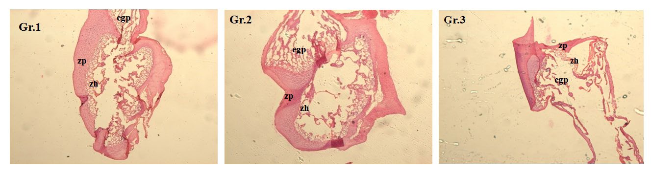
Figure 7: Histology of femur of pups on day 21 of lactation period (H-E counterstaining, 40X magnification). Zp= zone of proliferation, zh=zone of hypertrophy, egp= epiphyseal growth plate. Gr. 1: pups of control group, Gr. 2: pups of pregnant female rats treated with BTE (50 mg /kg body weight/day, p.o.), Gr.3: pups of pregnant female rats treated with BTE (100 mg /kg body weight/day, p.o.).
Discussion
Preeclampsia is linked to alterations of placental function leading to stress and apoptotic signalling which can lead to defects in the offspring. Apoptotic and stress signalling are augmented in preeclampsia placenta and cord tissue that alter the intrauterine environment and activates the detrimental signaling that is transported to fetus [30]. In the present study, IHC of placenta and uteri showed BTE (100 mg BTE/kg body weight/day, p.o.) increased the expression of Bax and caspase-3 which may be an indication of preeclampsia. BTE (100 mg BTE/kg body weight/day, p.o.) also increased the Bax/Bcl-2 ratio compared to control group.
Matrix metalloproteinase is a family of proteolytic enzymes, able to degrade extracellular matrix & basement membrane components. Activity and localization of MMP2 and 9 in vitro systems (tissue culture and amniotic fluid) were examined in several studies. Omran, et al. (2011) found strong MMP-2 protein expression in the majority of the preeclamptic placentas. MMP-2 also restricted intrauterine growth which may lead to remarkable low mean birth weight in patients with preeclampsia [31]. It was found that before the appearance of clinical symptoms, the plasma concentrations of MMP-2 was elevated in preeclamptic women. MMP-2 may play a role in causing hypertension during pregnancy through multiple complex pathways [32]. On the other hand, Bone Morphogenetic Protein 7 (BMP-7) is the member of the transforming growth factor β superfamily. In an experiment, BMP-7 was found to be a potent apoptotic signals for the undifferentiated limb mesoderm which was a potent inhibitory factor for joint formation exhibit an intense expression in the perichondrium of the developing cartilages [33]. BMP7 is an important factor during the process of implantation that contributes to healthy embryonic development [34]. The primary receptor for vascular endothelial growth factor (VEGF) is vascular endothelial growth factor receptor 2 (VEGFR-2), a crucial receptor involved in normal endothelial function. Significantly decrease in VEGFR-2 transcript and protein levels were found in preeclamptic placentae. Nevo, et al. (2013) showed a novel hypoxia-induced and preeclampsia-related down-regulation of VEGFR-2 in the human placenta. In hypoxic conditions and preeclampsia, sFlt-1 (Soluble fms-like tyrosine kinase-1) was found to be increased by which the VEGFR-2 expression and signaling get attenuated. A direct interaction between sFlt-1 and VEGFR-2 was involved in VEGFR-2 regulation, inhibition of VEGFR-2-mediated processes during placentation and also a novel platform to examine the onset of preeclampsia [35]. In the present study we found, BTE (100 mg /kg body weight/day, p.o.) significantly (p<0.05) increased MMP2+ placental cells but MMP2+VEGFR2+ and BMP7+ cell populations were decreased compared to control group. So it was clear that BTE (100 mg /kg body weight/day, p.o.) induced higher expression of MMP-2 and decreased level of BMP-7 causing preeclampsia and restriction in intrauterine growth. There was a further trigger of the apoptotic signal which might be responsible for increased the expression of Bax and caspase-3 and also increased the Bax/Bcl-2 ratio in BTE (100 mg /kg body weight/day, p.o.) treated rats compared to control group.
It was established the principal caffeine metabolism enzyme, cytochrome CYP1A2, is absent in the placenta and fetus [36]. Caffeine which is also a constituent of tea can freely passes through the placental barrier from the mother to the fetus and hence, maternal caffeine intake during pregnancy directly influences fetal caffeine exposure levels [37, 38]. Fetal exposure to caffeine increases circulating catecholamine concentrations, which might subsequently lead to fetoplacental vasoconstriction and hypoxia [19] and eventually affect fetal growth and development [40, 41, 42]. In a study, pu-erh black tea a highly fermented version of black tea is associated with development of fetal toxicity at a high concentration [19]. Though in another study there was no heat-sterilized green tea catechins (GTC-H) related fetal malformations or developmental variations [42].
Dey, et al. biomorphometric parameters like cranial length, cranial diameter, neck width, craniosacral length and tail length showed BTE retards the growth of pups but no significant changes were found in terms of time taken to open eyes, eruption of incisors and appearance of fur [22]. In present study, it was found that BTE decreased the bone mineral density of fetus and pups. It was clearly evident that BTE significantly decreased the concentration of Ca2+, P, Mg2+ and Zn2+ in bone in dose dependent manner and decreased the rate of ossification. BTE (100 mg BTE/kg body weight/ day, p.o.) remarkably altered the normal growth of bone as the zone of proliferation (zp) region and zone of hypertrophy (zh) with reduced epiphyseal growth plate (egp) in the femur of pups were noticed. These findings also supported the proposed findings. The animal results are also applicable to human. This study is the first time effort to establish the effect of Black tea extract at high dose on mother and pups during pregnancy and lactation period. Till now it is not clear which active component(s) of black tea is/are responsible for this negative action. On the other hand black tea constituents vary with internal and external factors. So different black tea varieties may affect prenatal and postnatal health in different level. The usage of different pesticides and herbicides in the tea gardens should not be ignored which may alter the health and hygiene of tea consumers. Further studies are warranted to establish the BTE induced changes at molecular level and physiological changes during pregnancy and development of embryos.
Conclusion
In conclusion, BTE increased the level of MMP-2, Bax and caspase-3 whereas decreased the level of BMP-7 in placenta which was an indication of preeclampsia. In fetus and pups, BTE significantly decreased the bone mineral density and the rate of ossification which were the outcomes of preeclampsia. This study confirmed BTE induced preeclampsia during pregnancy retarded the bone growth of fetus and pups in experimental animal model.
Acknowledgements
Authors would like to thank the Post Graduate Department of Zoology, Maulana Azad College, Kolkata for instruments facility and Department of Zoology and West Bengal State University for assisting in flow cytometry and its data analysis. Authors also thankful to the National Tea Research Foundation, Tea Board, India for financial assistance to carry out the present investigation. This research work was supported by the National Tea Research Foundation, Tea Board, India [Code No. NTRF: 164/2014; Ref No. NTRF: 17(305)/2013/4423 dated 11th March, 2014].
Conflict of Interests
The authors declare there is no conflict of interests exists.
References
-
Balentine DA, Harbowy ME, Graham HN (1998) Tea: The plant and its manufacture; chemistry and consumption of the Beverage. In: Spiller GA (Eds), Caffeine. Boca Raton, CRC Press, pp: 37-68.
-
Modder WWD, Amarakoon AMT (2002) Tea and Health. Tea Research Institute, Talawakelle, Sri Lanka, pp: 1-179.
-
Skotnicka M, Chorostowska-Wynimko J, Jankun J, Skrzypczak-Jankun E (2011) The Black tea bioactivity: an overview. Cent Eur J Immunol 36(4): 284-292.
-
Graham HN (1992) Green tea composition, consumption, and polyphenol chemistry. Prev Med 21(3): 334-350.
-
Wong CC, Cheng K, Chao J, Peng X, Zheng Z, et al. (2009) Analytical methods for bioactive compounds in teas. In: Ho CT, ett al. (Eds.), Tea and tea products: Chemistry and health-promoting properties. Boca Raton, CRC Press, pp: 77-110.
-
Hara Y (2001) Green tea: Health benefits and applications. Marcel Dekker Inc, New York.
-
Wan X, Li D, Zhang Z (2009) Green tea and black tea manufacturing and consumption. In: Ho CT, et al. (Eds.), Tea and tea products: Chemistry and health promoting properties. Boca Raton, CRC Press, pp: 1-8.
-
Astill C, Birch MR, Dacombe C, Humphrey PG, Martin PT (2001) Factors affecting the caffeine and polyphenol contents of black and green tea infusions. J Agric Food Chem 49(11): 5340-5347.
-
Jochmann N, Lorenz M, Krosigk A, Martus P, Bohm V, et al. (2008) The efficacy of black tea in ameliorating endothelial function is equivalent to that of green tea. Br J Nutr 99(4): 863-868.
-
Singh DK, Banerjee S, Porter TD (2009) Green and black tea extracts inhibit HMG-CoA reductase and activate AMP kinase to decrease cholesterol synthesis in hepatoma cells. J Nutr Biochem 20(10): 816-822.
-
Roy DK, Kumar KT, Karmakar S, Pal S, Samanta SK, et al. (2008) Pharmacological studies on Indian black tea (leaf variety) in acute and chronic inflammatory conditions. Phytother Res 22(6): 814-819.
-
de Mejia EG, Ramirez-Mares MV, Puangpraphant S (2009) Bioactive components of tea: cancer, inflammation and behaviour. Brain Behav Immun 23(6): 721-731.
-
Larsen CA, Dashwood RH, Bisson WH (2010) Tea catechins as inhibitors of receptor tyrosine kinases: mechanistic insights and human relevance. Pharmacol Res 62(6): 457-464.
-
Shih LJ, Lin YR, Lin CK, Liu HS, Kao YH (2016) Green tea (-)-epigallocatechin gallate induced growth inhibition of human placental choriocarcinoma cells. Placenta 41: 1-9.
-
Grove KA, Lambert JD (2010) Laboratory, Epidemiological, and Human Intervention Studies Show That Tea (Camellia sinensis) May Be Useful in the Prevention of Obesity. J Nutr 140(3): 446-453.
-
Skrzypczak-Jankun E, Jankun J (2010) Theaflavin digallate inactivates plasminogen activator inhibitor: Could tea help in Alzheimer’s disease and obesity? Int J Mol Med 26(1): 45-50.
-
Isbrucker RA, Edwards JA, Wolz E, Davidovich A, Bausch J (2006) Safety studies on epigallocatechin gallate (EGCG) preparations. Part 3: teratogenicity and reproductive toxicity studies in rats. Food Chem Toxicol 44(5): 651- 661.
-
Fan YC, Chan WH (2014) Epigallocatechin gallate induces embryonic toxicity in mouse blastocysts through apoptosis. Drug Chem Toxicol 37(3): 247-254.
-
Wang D, Meng J, Xu K, Xiao R, Xu M, et al. (2012) Evaluation of oral subchronic toxicity of Pu-erh green tea (Camellia sinensis var. assamica) extract in Sprague Dawley rats. J Ethnopharmacol 142(3): 836-844.
-
Saudan P, Brown MA, Buddle ML, Jones M (1998) Does gestational hypertension become pre-eclampsia? Br J Obstet Gynaecol 105(11): 1077-1084.
-
Hennessy A, Pilmore HL, Simmons LA, Painter DM (1999) A Deficiency of Placental IL-10 in Preeclampsia. J Immunol 163(6): 3491-3495.
-
Dey A, Gomes A, Dasgupta SC (2018) Black Tea (Camellia sinensis) Extract Induced Prenatal and Postnatal Toxicity in Experimental Albino rats. Pharmacogn Mag 13(Suppl 4): S769-S774.
-
Gekas C, Rhodes KE, Mikkola HKA (2008) Isolation and Analysis of Hematopoietic Stem Cells from the Placenta. J Vis Exp 16: 742.
-
Chakraborty K, Chatterjee S, Bhattacharyya A (2017) Modulation of CD11c+ lung dendritic cells in respect to TGF-β in experimental pulmonary fibrosis. Cell Biol Int 41(9): 991-1000.
-
Jungbluth AA, Stockert E, Huang HJS, Collins VP, Coplan K, et al. (2003) A monoclonal antibody recognizing human cancers with amplification/overexpression of the human epidermal growth factor receptor. Proc Natl Acad Sci 100(2): 639-644.
-
Ataee R, Ajdary S, Zarrindast M, Rezayat M, Hayatbakhsh MR (2010) Anti-mitogenic and apoptotic effects of 5-HT1B receptor antagonist on HT29 colorectal cancer cell line. J Cancer Res Clin Oncol 136(10): 1461-1469.
-
Menegola E, Broccia ML, Giavini E (2001) Atlas of Rat Fetal Skeleton Double Stained for Bone and Cartilage. Teratology 64(3): 125-133.
-
Gomes A, Haldar S, Giri B, Mishra R, Saha A, et al. (2009) Experimental osteoporosis induced in female albino rats and its antagonism by Indian black scorpion (Heterometrus bengalensis C.L.Koch) venom. Toxicon 53(1): 60-68.
-
Nunes JS (2016) Musculoskeletal System. In: Parker GA, Picut CA (Eds.) Atlas of Histology of Juvenile Rat 1st (Edn.), Academic Press, Cambridge, pp: 29-43.
-
Afroze SH, Kalagiri RR, Reyes M, Zimmerman JD, Beeram MR, et al. (2016) Apoptotic and stress signaling markers are augmented in preeclamptic placenta and umbilical cord. BBA Clin 6: 25-30.
-
Omran OM, Shokry M, Ismail H, Omar G, Rezk M (2011) Expression of matrix metalloproteinases 2 and 9 in human trophoblasts of normal and preeclamptic placentas. Int J Health Sci 5(2 Suppl 1): 21-23.
-
Palei ACT, Granger JP, Tanus-Santos JE (2013) Matrix Metalloproteinases as Drug Targets in Preeclampsia. Cur Drug Targets 14(3): 325-334.
-
Macias D, Ganan Y, Sampath TK, Piedra ME, Ros MA, et al. (1997) Role of BMP-2 and OP-1 (BMP-7) in programmed cell death and skeletogenesis during chick limb development. Development 124(6): 1109-1117.
-
Monsivais D, Clementi C, Peng J, Fullerton PT, Prunskaite- Hyyrylainen R, et al. (2017) BMP7 Induces Uterine Receptivity and Blastocyst Attachment. Endocrinology 158(4): 979-992.
-
Nevo O, Lee DK, Caniggia I (2013) Attenuation of VEGFR-2 Expression by sFlt-1 and Low Oxygen in Human Placenta. Plos One 8(11).
-
Aldridge A, Aranda JV, Neims AH (1979) Caffeine metabolism in the newborn. Clin Pharmacol Ther 25(4): 447-453.
-
Weathersbee PS, Lodge JR (1977) Caffeine: its direct and indirect influence on reproduction. J Reprod Med 19(2): 55-63.
-
Arnaud MJ, Bracco I, Sauvageat JL, Clerc MF (1983) Placental transfer of the major caffeine metabolite in the rat using 6-amino-5[N-formylmethylamino]1,3[Me- 14C]-dimethyluracil administered orally or intravenously to the pregnant rat. Toxicol Lett 16(3-4): 271-279.
-
Kirkinen P, Jouppila P, Koivula A, Vuori J, Puukka M (1983) The effect of caffeine on placental and fetal blood flow in human pregnancy. Am J Obstet Gynecol 147(8): 939-942.
-
Jarosz M, Wierzejska R, Siuba M (2012) Maternal caffeine intake and its effect on pregnancy outcomes. Eur J Obstet Gynecol Reprod Biol 160(2): 156-160.
-
Rhee J, Kim R, Kim Y, Tam M, Lai Y, et al. (2015) Maternal Caffeine Consumption during Pregnancy and Risk of Low Birth Weight: A Dose-Response Meta-Analysis of Observational Studies. PLOS ONE 10(7).
-
Morita O, Knapp JF, Tamaki Y, Stump DG, Moore JS, et al. (2009) Effects of green tea catechin on embryo/fetal development in rats. Food Chem Toxicol 47(6): 1296- 1303.
- Pattern of Gonadal Hormones in Oral Testosterone-Supplimented Male Wistar Rats with Diabetes-Induced Hypogonadism
- Re-Evaluation of the Genotoxicity of Currently Used Food Dyes in Mouse Multiple Organs Via Continuous Administration by Drinking Using the Comet Assay
- Pharmacogenetics of Type 2 Diabetes Mellitus: Linking Genetic Variability to Drug Efficacy and its Cardiovascular Outcomes
- Exploratory Proteomic Profiling of SARS-CoV-2 Infected THP-1 Macrophages Reveals Alterations in Inflammatory Response and Cellular Metabolism
- Study of Genotoxicity of Hepatocarcinogens in Multiple Organs in Mice by Feeding and Drinking Using the Comet Assay
- Spirulina Polypeptides Inhibit the Growth of Human Lung Tumor (H460) Cells